Structural characterization and functional analysis of cystathionine beta-synthase: an enzyme involved in the reverse transsulfuration pathway of Bacillus anthracis.
Devi, S., Abdul Rehman, S.A., Tarique, K.F., Gourinath, S.(2017) FEBS J 284: 3862-3880
- PubMed: 28921884
- DOI: https://doi.org/10.1111/febs.14273
- Primary Citation of Related Structures:
5XW3 - PubMed Abstract:
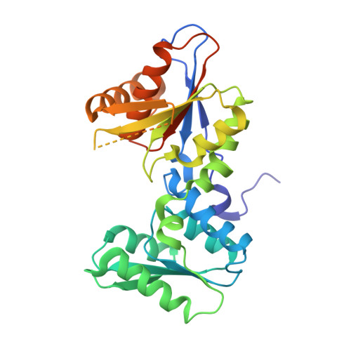
The reverse transsulfuration pathway has been reported to produce cysteine from homocysteine in eukaryotes ranging from protozoans to mammals while bacteria and plants produce cysteine via a de novo pathway. Interestingly, the bacterium Bacillus anthracis includes enzymes of the reverse transsulfuration pathway viz. cystathionine β-synthase [BaCBS, previously annotated to be an O-acetylserine sulfhydrylase (OASS)] and cystathionine γ-lyase. Here, we report the structure of BaCBS at a resolution of 2.2 Å. The enzyme was found to show CBS activity only with activated serine (O-acetylserine) and not with serine, and was also observed to display OASS activity but not serine sulfhydrylase activity. BaCBS was also found to produce hydrogen sulfide (H 2 S) upon reaction of cysteine and homocysteine. A mutational study revealed Glu 220, conserved in CBS, to be necessary for generating H 2 S. Structurally, BaCBS display a considerably more open active site than has been found for any other CBS or OASS, which was attributed to the presence of a helix at the junction of the C- and N-terminal domains. The root-mean-square deviation (RMSD) between the backbone Cα carbon atoms of BaCBS and those of other CBSs and OASSs were calculated to be greater than 3.0 Å. The pyridoxal 5'-phosphate at the active site was not traced, and appeared to be highly flexible due to the active site being wide open. Phylogenetic analysis revealed the presence of an O-acetylserine-dependent CBS in the bacterial domain and making separate clade from CBS and OASS indicating its evolution for specific function. Structural data are available in the PDB under the accession number 5XW3.
Organizational Affiliation:
Structural Biology Laboratory, School of Life Sciences, Jawaharlal Nehru University, New Delhi, India.