5XPF

High-resolution X-ray structure of the T26H mutant of T4 lysozyme


Experimental Data Snapshot

  • Method: X-RAY DIFFRACTION
  • Resolution: 1.04 Å
  • R-Value Free: 0.193 
  • R-Value Work: 0.159 

wwPDB Validation   3D Report Full Report


This is version 2.2 of the entry. See complete history


Literature

Neutron structure of the T26H mutant of T4 phage lysozyme provides insight into the catalytic activity of the mutant enzyme and how it differs from that of wild type.

Hiromoto, T.Meilleur, F.Shimizu, R.Shibazaki, C.Adachi, M.Tamada, T.Kuroki, R.

(2017) Protein Sci 26: 1953-1963

  • DOI: https://doi.org/10.1002/pro.3230
  • Primary Citation of Related Structures:  
    5XPE, 5XPF

  • PubMed Abstract: 

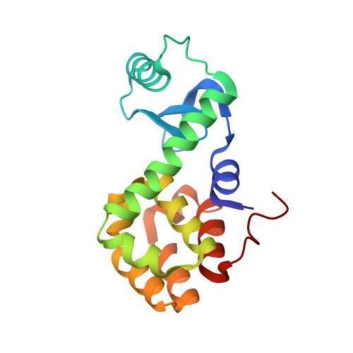
    T4 phage lysozyme is an inverting glycoside hydrolase that degrades the murein of bacterial cell walls by cleaving the β-1,4-glycosidic bond. The substitution of the catalytic Thr26 residue to a histidine converts the wild type from an inverting to a retaining enzyme, which implies that the original general acid Glu11 can also act as an acid/base catalyst in the hydrolysis. Here, we have determined the neutron structure of the perdeuterated T26H mutant to clarify the protonation states of Glu11 and the substituted His26, which are key in the retaining reaction. The 2.09-Å resolution structure shows that the imidazole group of His26 is in its singly protonated form in the active site, suggesting that the deprotonated Nɛ2 atom of His26 can attack the anomeric carbon of bound substrate as a nucleophile. The carboxyl group of Glu11 is partially protonated and interacts with the unusual neutral state of the guanidine moiety of Arg145, as well as two heavy water molecules. Considering that one of the water-binding sites has the potential to be occupied by a hydronium ion, the bulk solvent could be the source for the protonation of Glu11. The respective protonation states of Glu11 and His26 are consistent with the bond lengths determined by an unrestrained refinement of the high-resolution X-ray structure of T26H at 1.04-Å resolution. The detail structural information, including the coordinates of the deuterium atoms in the active site, provides insight into the distinctively different catalytic activities of the mutant and wild type enzymes.


  • Organizational Affiliation

    Quantum Beam Science Center, Japan Atomic Energy Agency, Tokai, Ibaraki, 319-1195, Japan.


Macromolecules
Find similar proteins by:  (by identity cutoff)  |  3D Structure
Entity ID: 1
MoleculeChains Sequence LengthOrganismDetailsImage
Endolysin164Tequatrovirus T4Mutation(s): 3 
Gene Names: eT4Tp126
EC: 3.2.1.17
UniProt
Find proteins for P00720 (Enterobacteria phage T4)
Explore P00720 
Go to UniProtKB:  P00720
Entity Groups  
Sequence Clusters30% Identity50% Identity70% Identity90% Identity95% Identity100% Identity
UniProt GroupP00720
Sequence Annotations
Expand
  • Reference Sequence
Small Molecules
Ligands 4 Unique
IDChains Name / Formula / InChI Key2D Diagram3D Interactions
HEZ
Query on HEZ

Download Ideal Coordinates CCD File 
J [auth A],
K [auth A],
L [auth A],
M [auth A]
HEXANE-1,6-DIOL
C6 H14 O2
XXMIOPMDWAUFGU-UHFFFAOYSA-N
GOL
Query on GOL

Download Ideal Coordinates CCD File 
N [auth A],
O [auth A],
P [auth A]
GLYCEROL
C3 H8 O3
PEDCQBHIVMGVHV-UHFFFAOYSA-N
CL
Query on CL

Download Ideal Coordinates CCD File 
E [auth A],
F [auth A],
G [auth A],
H [auth A],
I [auth A]
CHLORIDE ION
Cl
VEXZGXHMUGYJMC-UHFFFAOYSA-M
NA
Query on NA

Download Ideal Coordinates CCD File 
B [auth A],
C [auth A],
D [auth A]
SODIUM ION
Na
FKNQFGJONOIPTF-UHFFFAOYSA-N
Experimental Data & Validation

Experimental Data

  • Method: X-RAY DIFFRACTION
  • Resolution: 1.04 Å
  • R-Value Free: 0.193 
  • R-Value Work: 0.159 
  • Space Group: P 32 2 1
Unit Cell:
Length ( Å )Angle ( ˚ )
a = 59.839α = 90
b = 59.839β = 90
c = 95.828γ = 120
Software Package:
Software NamePurpose
HKL-2000data processing
HKL-2000data scaling
PDB_EXTRACTdata extraction
SHELXLrefinement
Cootmodel building

Structure Validation

View Full Validation Report


Entry History 

Deposition Data

Revision History  (Full details and data files)

  • Version 1.0: 2017-10-04
    Type: Initial release
  • Version 2.0: 2017-10-11
    Changes: Polymer sequence
  • Version 2.1: 2017-11-15
    Changes: Refinement description
  • Version 2.2: 2023-11-22
    Changes: Data collection, Database references, Derived calculations, Refinement description