5XOJ

Crystal structure of Xpo1p-PKI-Nup42p-Gsp1p-GTP complex


Experimental Data Snapshot

  • Method: X-RAY DIFFRACTION
  • Resolution: 2.20 Å
  • R-Value Free: 0.220 
  • R-Value Work: 0.191 
  • R-Value Observed: 0.192 

wwPDB Validation   3D Report Full Report


Ligand Structure Quality Assessment 


This is version 1.2 of the entry. See complete history


Literature

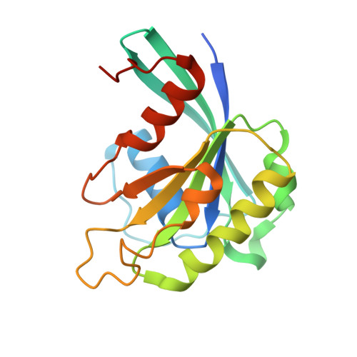
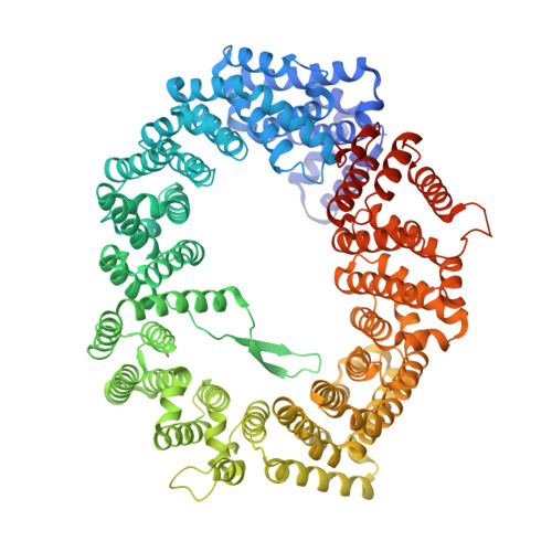
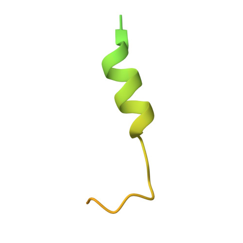
Crystal structure of the Xpo1p nuclear export complex bound to the SxFG/PxFG repeats of the nucleoporin Nup42p

Koyama, M.Hirano, H.Shirai, N.Matsuura, Y.

(2017) Genes Cells 22: 861-875

  • DOI: https://doi.org/10.1111/gtc.12520
  • Primary Citation of Related Structures:  
    5XOJ

  • PubMed Abstract: 

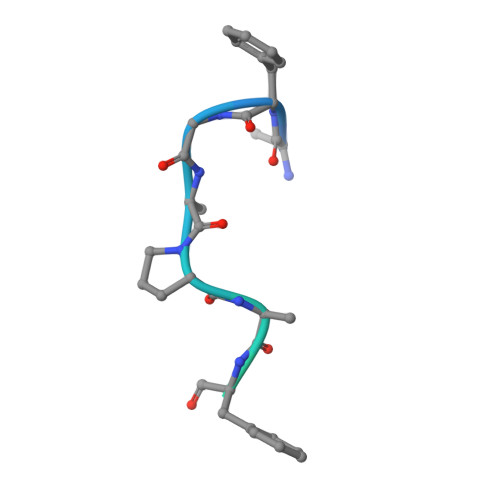
    Xpo1p (yeast CRM1) is the major nuclear export receptor that carries a plethora of proteins and ribonucleoproteins from the nucleus to cytoplasm. The passage of the Xpo1p nuclear export complex through nuclear pore complexes (NPCs) is facilitated by interactions with nucleoporins (Nups) containing extensive repeats of phenylalanine-glycine (so-called FG repeats), although the precise role of each Nup in the nuclear export reaction remains incompletely understood. Here we report structural and biochemical characterization of the interactions between the Xpo1p nuclear export complex and the FG repeats of Nup42p, a nucleoporin localized at the cytoplasmic face of yeast NPCs and has characteristic SxFG/PxFG sequence repeat motif. The crystal structure of Xpo1p-PKI-Nup42p-Gsp1p-GTP complex identified three binding sites for the SxFG/PxFG repeats on HEAT repeats 14-20 of Xpo1p. Mutational analyses of Nup42p showed that the conserved serines and prolines in the SxFG/PxFG repeats contribute to Xpo1p-Nup42p binding. Our structural and biochemical data suggest that SxFG/PxFG-Nups such as Nup42p and Nup159p at the cytoplasmic face of NPCs provide high-affinity docking sites for the Xpo1p nuclear export complex in the terminal stage of NPC passage and that subsequent disassembly of the nuclear export complex facilitates recycling of free Xpo1p back to the nucleus.


  • Organizational Affiliation

    Division of Biological Science, Nagoya University, Nagoya, 464-8602, Japan.


Macromolecules
Find similar proteins by:  (by identity cutoff)  |  3D Structure
Entity ID: 1
MoleculeChains Sequence LengthOrganismDetailsImage
GTP-binding nuclear protein182Saccharomyces cerevisiae AWRI796Mutation(s): 1 
Gene Names: AWRI796_3356
Entity Groups  
Sequence Clusters30% Identity50% Identity70% Identity90% Identity95% Identity100% Identity
Sequence Annotations
Expand
  • Reference Sequence
Find similar proteins by:  (by identity cutoff)  |  3D Structure
Entity ID: 2
MoleculeChains Sequence LengthOrganismDetailsImage
Exportin-1B [auth C]1,047Saccharomyces cerevisiae S288CMutation(s): 0 
Gene Names: CRM1KAP124XPO1YGR218WG8514
UniProt
Find proteins for P30822 (Saccharomyces cerevisiae (strain ATCC 204508 / S288c))
Explore P30822 
Go to UniProtKB:  P30822
Entity Groups  
Sequence Clusters30% Identity50% Identity70% Identity90% Identity95% Identity100% Identity
UniProt GroupP30822
Sequence Annotations
Expand
  • Reference Sequence
Find similar proteins by:  (by identity cutoff)  |  3D Structure
Entity ID: 3
MoleculeChains Sequence LengthOrganismDetailsImage
cAMP-dependent protein kinase inhibitor alphaC [auth D]76Homo sapiensMutation(s): 1 
Gene Names: PKIAPRKACN1
UniProt & NIH Common Fund Data Resources
Find proteins for P61925 (Homo sapiens)
Explore P61925 
Go to UniProtKB:  P61925
PHAROS:  P61925
GTEx:  ENSG00000171033 
Entity Groups  
Sequence Clusters30% Identity50% Identity70% Identity90% Identity95% Identity100% Identity
UniProt GroupP61925
Sequence Annotations
Expand
  • Reference Sequence
Find similar proteins by:  (by identity cutoff)  |  3D Structure
Entity ID: 4
MoleculeChains Sequence LengthOrganismDetailsImage
Nup42pD [auth E],
E [auth F],
F [auth G]
35Saccharomyces cerevisiae FostersBMutation(s): 0 
Entity Groups  
Sequence Clusters30% Identity50% Identity70% Identity90% Identity95% Identity100% Identity
Sequence Annotations
Expand
  • Reference Sequence
Small Molecules
Ligands 2 Unique
IDChains Name / Formula / InChI Key2D Diagram3D Interactions
GTP
Query on GTP

Download Ideal Coordinates CCD File 
G [auth A]GUANOSINE-5'-TRIPHOSPHATE
C10 H16 N5 O14 P3
XKMLYUALXHKNFT-UUOKFMHZSA-N
MG
Query on MG

Download Ideal Coordinates CCD File 
H [auth A]MAGNESIUM ION
Mg
JLVVSXFLKOJNIY-UHFFFAOYSA-N
Experimental Data & Validation

Experimental Data

  • Method: X-RAY DIFFRACTION
  • Resolution: 2.20 Å
  • R-Value Free: 0.220 
  • R-Value Work: 0.191 
  • R-Value Observed: 0.192 
  • Space Group: P 21 21 21
Unit Cell:
Length ( Å )Angle ( ˚ )
a = 101.64α = 90
b = 107.56β = 90
c = 149.11γ = 90
Software Package:
Software NamePurpose
Aimlessdata scaling
PHENIXrefinement
PDB_EXTRACTdata extraction
iMOSFLMdata reduction
MOLREPphasing

Structure Validation

View Full Validation Report


Ligand Structure Quality Assessment 


Entry History 

Deposition Data

Revision History  (Full details and data files)

  • Version 1.0: 2017-08-16
    Type: Initial release
  • Version 1.1: 2017-11-01
    Changes: Database references
  • Version 1.2: 2023-11-22
    Changes: Data collection, Database references, Derived calculations, Refinement description