5X3T

VapBC from Mycobacterium tuberculosis


Experimental Data Snapshot

  • Method: X-RAY DIFFRACTION
  • Resolution: 2.65 Å
  • R-Value Free: 0.239 
  • R-Value Work: 0.208 
  • R-Value Observed: 0.210 

wwPDB Validation   3D Report Full Report


This is version 1.1 of the entry. See complete history


Literature

Functional details of the Mycobacterium tuberculosis VapBC26 toxin-antitoxin system based on a structural study: insights into unique binding and antibiotic peptides.

Kang, S.M.Kim, D.H.Lee, K.Y.Park, S.J.Yoon, H.J.Lee, S.J.Im, H.Lee, B.J.

(2017) Nucleic Acids Res 45: 8564-8580

  • DOI: https://doi.org/10.1093/nar/gkx489
  • Primary Citation of Related Structures:  
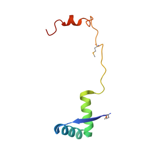
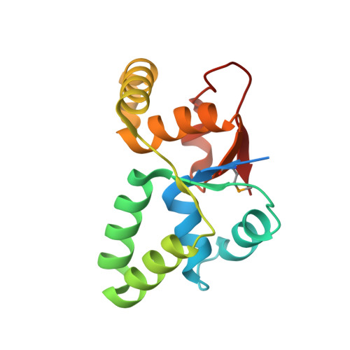
    5X3T

  • PubMed Abstract: 

    Toxin-antitoxin (TA) systems are essential for bacterial persistence under stressful conditions. In particular, Mycobacterium tuberculosis express VapBC TA genes that encode the stable VapC toxin and the labile VapB antitoxin. Under normal conditions, these proteins interact to form a non-toxic TA complex, but the toxin is activated by release from the antitoxin in response to unfavorable conditions. Here, we present the crystal structure of the M. tuberculosis VapBC26 complex and show that the VapC26 toxin contains a pilus retraction protein (PilT) N-terminal (PIN) domain that is essential for ribonuclease activity and that, the VapB26 antitoxin folds into a ribbon-helix-helix DNA-binding motif at the N-terminus. The active site of VapC26 is sterically blocked by the flexible C-terminal region of VapB26. The C-terminal region of free VapB26 adopts an unfolded conformation but forms a helix upon binding to VapC26. The results of RNase activity assays show that Mg2+ and Mn2+ are essential for the ribonuclease activity of VapC26. As shown in the nuclear magnetic resonance spectra, several residues of VapB26 participate in the specific binding to the promoter region of the VapBC26 operon. In addition, toxin-mimicking peptides were designed that inhibit TA complex formation and thereby increase toxin activity, providing a novel approach to the development of new antibiotics.


  • Organizational Affiliation

    The Research Institute of Pharmaceutical Sciences, College of Pharmacy, Seoul National University, Gwanak-gu, Seoul 151-742, Republic of Korea.


Macromolecules
Find similar proteins by:  (by identity cutoff)  |  3D Structure
Entity ID: 1
MoleculeChains Sequence LengthOrganismDetailsImage
Antitoxin VapB26A,
B [auth C],
C [auth E],
D [auth G]
71Mycobacterium tuberculosis H37RvMutation(s): 1 
Gene Names: vapB26Rv0581
UniProt
Find proteins for O53778 (Mycobacterium tuberculosis (strain ATCC 25618 / H37Rv))
Explore O53778 
Go to UniProtKB:  O53778
Entity Groups  
Sequence Clusters30% Identity50% Identity70% Identity90% Identity95% Identity100% Identity
UniProt GroupO53778
Sequence Annotations
Expand
  • Reference Sequence
Find similar proteins by:  (by identity cutoff)  |  3D Structure
Entity ID: 2
MoleculeChains Sequence LengthOrganismDetailsImage
Ribonuclease VapC26E [auth B],
F [auth D],
G [auth F],
H
155Mycobacterium tuberculosis H37RvMutation(s): 0 
Gene Names: vapC26Rv0582
EC: 3.1
UniProt
Find proteins for O53779 (Mycobacterium tuberculosis (strain ATCC 25618 / H37Rv))
Explore O53779 
Go to UniProtKB:  O53779
Entity Groups  
Sequence Clusters30% Identity50% Identity70% Identity90% Identity95% Identity100% Identity
UniProt GroupO53779
Sequence Annotations
Expand
  • Reference Sequence
Small Molecules
Ligands 1 Unique
IDChains Name / Formula / InChI Key2D Diagram3D Interactions
MG
Query on MG

Download Ideal Coordinates CCD File 
I [auth H]MAGNESIUM ION
Mg
JLVVSXFLKOJNIY-UHFFFAOYSA-N
Modified Residues  1 Unique
IDChains TypeFormula2D DiagramParent
MSE
Query on MSE
A,
B [auth C],
C [auth E],
D [auth G]
L-PEPTIDE LINKINGC5 H11 N O2 SeMET
Experimental Data & Validation

Experimental Data

  • Method: X-RAY DIFFRACTION
  • Resolution: 2.65 Å
  • R-Value Free: 0.239 
  • R-Value Work: 0.208 
  • R-Value Observed: 0.210 
  • Space Group: P 41
Unit Cell:
Length ( Å )Angle ( ˚ )
a = 64.221α = 90
b = 64.221β = 90
c = 216.134γ = 90
Software Package:
Software NamePurpose
PHENIXrefinement
HKL-2000data reduction
HKL-2000data scaling
AutoSolphasing

Structure Validation

View Full Validation Report


Entry History 

Deposition Data

Revision History  (Full details and data files)

  • Version 1.0: 2017-06-07
    Type: Initial release
  • Version 1.1: 2017-12-06
    Changes: Database references