5WVN

Crystal structure of MBS-BaeS fusion protein


Experimental Data Snapshot

  • Method: X-RAY DIFFRACTION
  • Resolution: 2.80 Å
  • R-Value Free: 0.270 
  • R-Value Work: 0.228 
  • R-Value Observed: 0.230 

wwPDB Validation   3D Report Full Report


This is version 1.2 of the entry. See complete history


Literature

Crystal structure of the sensor domain of BaeS from Serratia marcescens FS14

Zhang, Y.Qiu, S.Jia, S.Xu, D.Ran, T.Wang, W.

(2017) Proteins 85: 1784-1790

  • DOI: https://doi.org/10.1002/prot.25326
  • Primary Citation of Related Structures:  
    5WVM, 5WVN

  • PubMed Abstract: 

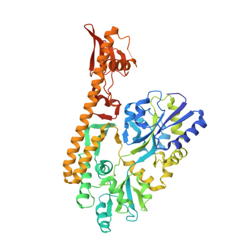
    The sensor histidine kinases of two-component signal-transduction systems (TCSs) are essential for bacteria to adapt to variable environmental conditions. The two-component regulatory system BaeS/R increases multidrug and metal resistance in Salmonella and Escherichia coli. In this study, we report the X-ray structure of the periplasmic sensor domain of BaeS from Serratia marcescens FS14. The BaeS sensor domain (34-160) adopts a mixed α/β-fold containing a central four-stranded antiparallel β-sheet flanked by a long N-terminal α-helix and additional loops and a short C-terminal α-helix on each side. Structural comparisons revealed that it belongs to the PDC family with a remarkable difference in the orientation of the helix α2. In the BaeS sensor domain, this helix is situated perpendicular to the long helix α1 and holds helix α1 in the middle with the beta sheet, whereas in other PDC domains, helix α2 is parallel to helix α1. Because the helices α1 and α2 is involved in the dimeric interface, this difference implies that BaeS uses a different dimeric interface compared with other PDC domains. Proteins 2017; 85:1784-1790. © 2017 Wiley Periodicals, Inc.


  • Organizational Affiliation

    Key Laboratory of Agricultural and Environmental Microbiology, Ministry of Agriculture, College of Life Sciences, Department of Microbiology, Nanjing Agricultural University, Nanjing, 210095, China.


Macromolecules
Find similar proteins by:  (by identity cutoff)  |  3D Structure
Entity ID: 1
MoleculeChains Sequence LengthOrganismDetailsImage
Maltose-binding periplasmic protein,Two-component system sensor kinase509Escherichia coli K-12Serratia marcescens subsp. marcescens Db11
This entity is chimeric
Mutation(s): 0 
Gene Names: malEb4034JW3994baeSSMDB11_2949
UniProt
Find proteins for P0AEX9 (Escherichia coli (strain K12))
Explore P0AEX9 
Go to UniProtKB:  P0AEX9
Entity Groups  
Sequence Clusters30% Identity50% Identity70% Identity90% Identity95% Identity100% Identity
UniProt GroupP0AEX9
Sequence Annotations
Expand
  • Reference Sequence
Experimental Data & Validation

Experimental Data

  • Method: X-RAY DIFFRACTION
  • Resolution: 2.80 Å
  • R-Value Free: 0.270 
  • R-Value Work: 0.228 
  • R-Value Observed: 0.230 
  • Space Group: P 43 21 2
Unit Cell:
Length ( Å )Angle ( ˚ )
a = 119.084α = 90
b = 119.084β = 90
c = 147.011γ = 90
Software Package:
Software NamePurpose
PHENIXrefinement
XDSdata reduction
SCALAdata scaling
PHASERphasing

Structure Validation

View Full Validation Report


Entry History & Funding Information

Deposition Data


Funding OrganizationLocationGrant Number
Natural Science Foundation of ChinaChina31170686
Natural Science Foundation of ChinaChina31400055
the Natural Science Foundation of Jiangsu ProvinceChinaBK20140690

Revision History  (Full details and data files)

  • Version 1.0: 2018-01-03
    Type: Initial release
  • Version 1.1: 2019-01-16
    Changes: Data collection, Database references
  • Version 1.2: 2024-03-20
    Changes: Data collection, Database references