5WL0

Co-crystal structure of Influenza A H3N2 PB2 (241-741) bound to VX-787


Experimental Data Snapshot

  • Method: X-RAY DIFFRACTION
  • Resolution: 2.40 Å
  • R-Value Free: 0.253 
  • R-Value Work: 0.203 
  • R-Value Observed: 0.205 

Starting Models: experimental
View more details

wwPDB Validation   3D Report Full Report


Ligand Structure Quality Assessment 


This is version 1.1 of the entry. See complete history


Literature

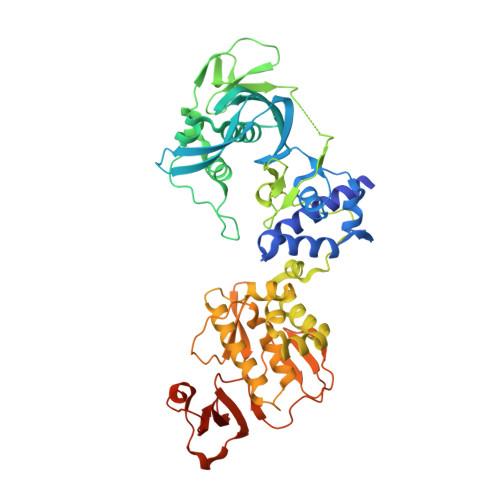
Structural basis for therapeutic inhibition of influenza A polymerase PB2 subunit.

Ma, X.Xie, L.Wartchow, C.Warne, R.Xu, Y.Rivkin, A.Tully, D.Shia, S.Uehara, K.Baldwin, D.M.Muiru, G.Zhong, W.Zaror, I.Bussiere, D.E.Leonard, V.H.J.

(2017) Sci Rep 7: 9385-9385

  • DOI: https://doi.org/10.1038/s41598-017-09538-x
  • Primary Citation of Related Structures:  
    5WL0

  • PubMed Abstract: 

    Influenza virus uses a unique mechanism to initiate viral transcription named cap-snatching. The PB2 subunit of the viral heterotrimeric RNA polymerase binds the cap structure of cellular pre-mRNA to promote its cleavage by the PA subunit. The resulting 11-13 capped oligomer is used by the PB1 polymerase subunit to initiate transcription of viral proteins. VX-787 is an inhibitor of the influenza A virus pre-mRNA cap-binding protein PB2. This clinical stage compound was shown to bind the minimal cap-binding domain of PB2 to inhibit the cap-snatching machinery. However, the binding of this molecule in the context of an extended form of the PB2 subunit has remained elusive. Here we generated a collection of PB2 truncations to identify a PB2 protein representative of its structure in the viral heterotrimeric protein. We present the crystal structure of VX-787 bound to a PB2 construct that recapitulates VX-787's biological antiviral activity in vitro. This co-structure reveals more extensive interactions than previously identified and provides insight into the observed resistance profile, affinity, binding kinetics, and conformational rearrangements induced by VX-787.


  • Organizational Affiliation

    Structural and Biophysical Chemistry, Novartis Institutes for BioMedical Research, Emeryville, CA, USA. xiaolei.ma@novartis.com.


Macromolecules
Find similar proteins by:  (by identity cutoff)  |  3D Structure
Entity ID: 1
MoleculeChains Sequence LengthOrganismDetailsImage
Polymerase basic protein 2510Influenza A virus (A/Udorn/307/1972(H3N2))Mutation(s): 0 
Gene Names: PB2
UniProt
Find proteins for Q67296 (Influenza A virus (strain A/Udorn/307/1972 H3N2))
Explore Q67296 
Go to UniProtKB:  Q67296
Entity Groups  
Sequence Clusters30% Identity50% Identity70% Identity90% Identity95% Identity100% Identity
UniProt GroupQ67296
Sequence Annotations
Expand
  • Reference Sequence
Small Molecules
Ligands 2 Unique
IDChains Name / Formula / InChI Key2D Diagram3D Interactions
21G
Query on 21G

Download Ideal Coordinates CCD File 
B [auth A](2S,3S)-3-[[5-fluoranyl-2-(5-fluoranyl-1H-pyrrolo[2,3-b]pyridin-3-yl)pyrimidin-4-yl]amino]bicyclo[2.2.2]octane-2-carboxylic acid
C20 H19 F2 N5 O2
JGPXDNKSIXAZEQ-UIHHKEIPSA-N
PEG
Query on PEG

Download Ideal Coordinates CCD File 
C [auth A]DI(HYDROXYETHYL)ETHER
C4 H10 O3
MTHSVFCYNBDYFN-UHFFFAOYSA-N
Binding Affinity Annotations 
IDSourceBinding Affinity
21G BindingDB:  5WL0 Kd: 54 (nM) from 1 assay(s)
EC50: 0.6 (nM) from 1 assay(s)
Experimental Data & Validation

Experimental Data

  • Method: X-RAY DIFFRACTION
  • Resolution: 2.40 Å
  • R-Value Free: 0.253 
  • R-Value Work: 0.203 
  • R-Value Observed: 0.205 
  • Space Group: C 1 2 1
Unit Cell:
Length ( Å )Angle ( ˚ )
a = 142.413α = 90
b = 56.752β = 111.23
c = 82.014γ = 90
Software Package:
Software NamePurpose
Aimlessdata scaling
PHASERphasing
PHENIXrefinement
PDB_EXTRACTdata extraction

Structure Validation

View Full Validation Report


Ligand Structure Quality Assessment 


Entry History 

Deposition Data

Revision History  (Full details and data files)

  • Version 1.0: 2017-09-06
    Type: Initial release
  • Version 1.1: 2023-10-04
    Changes: Data collection, Database references, Refinement description, Structure summary