5WHG

Vms1 mitochondrial localization core


Experimental Data Snapshot

  • Method: X-RAY DIFFRACTION
  • Resolution: 2.70 Å
  • R-Value Free: 0.245 
  • R-Value Work: 0.187 
  • R-Value Observed: 0.193 

wwPDB Validation   3D Report Full Report


This is version 1.2 of the entry. See complete history


Literature

Sterol Oxidation Mediates Stress-Responsive Vms1 Translocation to Mitochondria.

Nielson, J.R.Fredrickson, E.K.Waller, T.C.Rendon, O.Z.Schubert, H.L.Lin, Z.Hill, C.P.Rutter, J.

(2017) Mol Cell 68: 673-685.e6

  • DOI: https://doi.org/10.1016/j.molcel.2017.10.022
  • Primary Citation of Related Structures:  
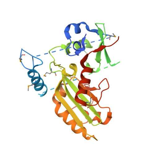
    5WHG

  • PubMed Abstract: 

    Vms1 translocates to damaged mitochondria in response to stress, whereupon its binding partner, Cdc48, contributes to mitochondrial protein homeostasis. Mitochondrial targeting of Vms1 is mediated by its conserved mitochondrial targeting domain (MTD), which, in unstressed conditions, is inhibited by intramolecular binding to the Vms1 leucine-rich sequence (LRS). Here, we report a 2.7 Å crystal structure of Vms1 that reveals that the LRS lies in a hydrophobic groove in the autoinhibited MTD. We also demonstrate that the oxidized sterol, ergosterol peroxide, is necessary and sufficient for Vms1 localization to mitochondria, through binding the MTD in an interaction that is competitive with binding to the LRS. These data support a model in which stressed mitochondria generate an oxidized sterol receptor that recruits Vms1 to support mitochondrial protein homeostasis.


  • Organizational Affiliation

    Department of Biochemistry, University of Utah School of Medicine, Salt Lake City, UT 84112, USA.


Macromolecules
Find similar proteins by:  (by identity cutoff)  |  3D Structure
Entity ID: 1
MoleculeChains Sequence LengthOrganismDetailsImage
Protein VMS1389Saccharomyces cerevisiae S288CMutation(s): 0 
Gene Names: VMS1YDR049W
UniProt
Find proteins for Q04311 (Saccharomyces cerevisiae (strain ATCC 204508 / S288c))
Explore Q04311 
Go to UniProtKB:  Q04311
Entity Groups  
Sequence Clusters30% Identity50% Identity70% Identity90% Identity95% Identity100% Identity
UniProt GroupQ04311
Sequence Annotations
Expand
  • Reference Sequence
Small Molecules
Ligands 1 Unique
IDChains Name / Formula / InChI Key2D Diagram3D Interactions
ZN
Query on ZN

Download Ideal Coordinates CCD File 
B [auth A]ZINC ION
Zn
PTFCDOFLOPIGGS-UHFFFAOYSA-N
Modified Residues  1 Unique
IDChains TypeFormula2D DiagramParent
MSE
Query on MSE
A
L-PEPTIDE LINKINGC5 H11 N O2 SeMET
Experimental Data & Validation

Experimental Data

  • Method: X-RAY DIFFRACTION
  • Resolution: 2.70 Å
  • R-Value Free: 0.245 
  • R-Value Work: 0.187 
  • R-Value Observed: 0.193 
  • Space Group: P 32 2 1
Unit Cell:
Length ( Å )Angle ( ˚ )
a = 62.864α = 90
b = 62.864β = 90
c = 154.383γ = 120
Software Package:
Software NamePurpose
SCALEPACKdata scaling
PHENIXrefinement
PDB_EXTRACTdata extraction
DENZOdata reduction
PHASERphasing

Structure Validation

View Full Validation Report


Entry History & Funding Information

Deposition Data


Funding OrganizationLocationGrant Number
National Institutes of Health/National Institute of General Medical Sciences (NIH/NIGMS)United StatesR01GM115129

Revision History  (Full details and data files)

  • Version 1.0: 2017-11-15
    Type: Initial release
  • Version 1.1: 2017-11-29
    Changes: Database references
  • Version 1.2: 2020-01-01
    Changes: Author supporting evidence