5VGR

Human Atlastin-3, GDP-bound


Experimental Data Snapshot

  • Method: X-RAY DIFFRACTION
  • Resolution: 2.10 Å
  • R-Value Free: 0.228 
  • R-Value Work: 0.198 
  • R-Value Observed: 0.199 

wwPDB Validation   3D Report Full Report


Ligand Structure Quality Assessment 


This is version 1.5 of the entry. See complete history


Literature

Timing and Reset Mechanism of GTP Hydrolysis-Driven Conformational Changes of Atlastin.

O'Donnell, J.P.Cooley, R.B.Kelly, C.M.Miller, K.Andersen, O.S.Rusinova, R.Sondermann, H.

(2017) Structure 25: 997-1010.e4

  • DOI: https://doi.org/10.1016/j.str.2017.05.007
  • Primary Citation of Related Structures:  
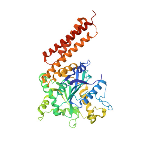
    5VGR

  • PubMed Abstract: 

    The endoplasmic reticulum (ER) forms a branched, dynamic membrane tubule network that is vital for cellular function. Branching arises from membrane fusion facilitated by the GTPase atlastin (ATL). Many metazoan genomes encode for three ATL isoforms that appear to fulfill partially redundant function despite differences in their intrinsic GTPase activity and localization within the ER; however, the underlying mechanistic differences between the isoforms are poorly understood. Here, we identify discrete temporal steps in the catalytic cycle for the two most dissimilar isoforms, ATL1 and ATL3, revealing an overall conserved progression of molecular events from nucleotide binding and hydrolysis to ATL dimerization and phosphate release. A crystal structure of ATL3 suggests a mechanism for the displacement of the catalytic Mg 2+ ion following guanosine triphosphate (GTP) hydrolysis. Together, the data extend the mechanistic framework for how GTP hydrolysis drives conformational changes in ATL and how the cycle is reset for subsequent rounds of catalysis.


  • Organizational Affiliation

    Department of Molecular Medicine, College of Veterinary Medicine, Cornell University, Ithaca, NY 14853, USA.


Macromolecules
Find similar proteins by:  (by identity cutoff)  |  3D Structure
Entity ID: 1
MoleculeChains Sequence LengthOrganismDetailsImage
Atlastin-3
A, B
431Homo sapiensMutation(s): 0 
Gene Names: ATL3
EC: 3.6.5
UniProt & NIH Common Fund Data Resources
Find proteins for Q6DD88 (Homo sapiens)
Explore Q6DD88 
Go to UniProtKB:  Q6DD88
PHAROS:  Q6DD88
GTEx:  ENSG00000184743 
Entity Groups  
Sequence Clusters30% Identity50% Identity70% Identity90% Identity95% Identity100% Identity
UniProt GroupQ6DD88
Sequence Annotations
Expand
  • Reference Sequence
Small Molecules
Experimental Data & Validation

Experimental Data

  • Method: X-RAY DIFFRACTION
  • Resolution: 2.10 Å
  • R-Value Free: 0.228 
  • R-Value Work: 0.198 
  • R-Value Observed: 0.199 
  • Space Group: C 1 2 1
Unit Cell:
Length ( Å )Angle ( ˚ )
a = 130.666α = 90
b = 57.057β = 116.22
c = 143.423γ = 90
Software Package:
Software NamePurpose
PHENIXrefinement
XDSdata reduction
SCALAdata scaling
PHENIXphasing

Structure Validation

View Full Validation Report


Ligand Structure Quality Assessment 


Entry History & Funding Information

Deposition Data


Funding OrganizationLocationGrant Number
National Institutes of Health/National Institute of General Medical Sciences (NIH/NIGMS)United StatesT32 GM008500

Revision History  (Full details and data files)

  • Version 1.0: 2017-05-17
    Type: Initial release
  • Version 1.1: 2017-06-28
    Changes: Database references
  • Version 1.2: 2017-07-19
    Changes: Database references
  • Version 1.3: 2017-09-20
    Changes: Author supporting evidence
  • Version 1.4: 2020-01-01
    Changes: Author supporting evidence
  • Version 1.5: 2023-10-04
    Changes: Data collection, Database references, Refinement description