5UWU

Crystal Structure of SMAD4 NES Peptide in complex with CRM1-Ran-RanBP1


Experimental Data Snapshot

  • Method: X-RAY DIFFRACTION
  • Resolution: 2.24 Å
  • R-Value Free: 0.211 
  • R-Value Work: 0.181 
  • R-Value Observed: 0.182 

wwPDB Validation   3D Report Full Report


Ligand Structure Quality Assessment 


This is version 1.4 of the entry. See complete history


Literature

Nuclear export receptor CRM1 recognizes diverse conformations in nuclear export signals.

Fung, H.Y.Fu, S.C.Chook, Y.M.

(2017) Elife 6

  • DOI: https://doi.org/10.7554/eLife.23961
  • Primary Citation of Related Structures:  
    5UWH, 5UWI, 5UWJ, 5UWO, 5UWP, 5UWQ, 5UWR, 5UWS, 5UWT, 5UWU, 5UWW

  • PubMed Abstract: 

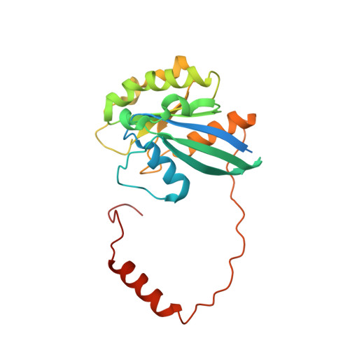
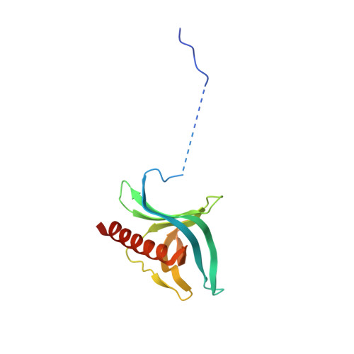
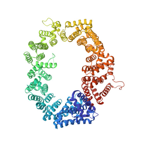
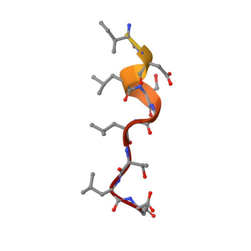
    Nuclear export receptor CRM1 binds highly variable nuclear export signals (NESs) in hundreds of different cargoes. Previously we have shown that CRM1 binds NESs in both polypeptide orientations (Fung et al., 2015). Here, we show crystal structures of CRM1 bound to eight additional NESs which reveal diverse conformations that range from loop-like to all-helix, which occupy different extents of the invariant NES-binding groove. Analysis of all NES structures show 5-6 distinct backbone conformations where the only conserved secondary structural element is one turn of helix that binds the central portion of the CRM1 groove. All NESs also participate in main chain hydrogen bonding with human CRM1 Lys568 side chain, which acts as a specificity filter that prevents binding of non-NES peptides. The large conformational range of NES backbones explains the lack of a fixed pattern for its 3-5 hydrophobic anchor residues, which in turn explains the large array of peptide sequences that can function as NESs.


  • Organizational Affiliation

    Department of Pharmacology, University of Texas Southwestern Medical Center, Dallas, United States.


Macromolecules
Find similar proteins by:  (by identity cutoff)  |  3D Structure
Entity ID: 1
MoleculeChains Sequence LengthOrganismDetailsImage
GTP-binding nuclear protein Ran237Homo sapiensMutation(s): 0 
Gene Names: RANARA24OK/SW-cl.81
UniProt & NIH Common Fund Data Resources
Find proteins for P62826 (Homo sapiens)
Explore P62826 
Go to UniProtKB:  P62826
PHAROS:  P62826
GTEx:  ENSG00000132341 
Entity Groups  
Sequence Clusters30% Identity50% Identity70% Identity90% Identity95% Identity100% Identity
UniProt GroupP62826
Sequence Annotations
Expand
  • Reference Sequence
Find similar proteins by:  (by identity cutoff)  |  3D Structure
Entity ID: 2
MoleculeChains Sequence LengthOrganismDetailsImage
Ran-specific GTPase-activating protein 1143Saccharomyces cerevisiaeMutation(s): 0 
Gene Names: YRB1CST20HTN1SFO1YDR002WYD8119.08
UniProt
Find proteins for P41920 (Saccharomyces cerevisiae (strain ATCC 204508 / S288c))
Explore P41920 
Go to UniProtKB:  P41920
Entity Groups  
Sequence Clusters30% Identity50% Identity70% Identity90% Identity95% Identity100% Identity
UniProt GroupP41920
Sequence Annotations
Expand
  • Reference Sequence
Find similar proteins by:  (by identity cutoff)  |  3D Structure
Entity ID: 3
MoleculeChains Sequence LengthOrganismDetailsImage
Exportin-11,024Saccharomyces cerevisiaeMutation(s): 0 
Gene Names: CRM1KAP124XPO1YGR218WG8514
UniProt
Find proteins for P30822 (Saccharomyces cerevisiae (strain ATCC 204508 / S288c))
Explore P30822 
Go to UniProtKB:  P30822
Entity Groups  
Sequence Clusters30% Identity50% Identity70% Identity90% Identity95% Identity100% Identity
UniProt GroupP30822
Sequence Annotations
Expand
  • Reference Sequence

Find similar proteins by:  Sequence   |   3D Structure  

Entity ID: 4
MoleculeChains Sequence LengthOrganismDetailsImage
Mothers against decapentaplegic homolog 420Homo sapiensMutation(s): 0 
Gene Names: SMAD4
UniProt & NIH Common Fund Data Resources
Find proteins for Q13485 (Homo sapiens)
Explore Q13485 
Go to UniProtKB:  Q13485
PHAROS:  Q13485
GTEx:  ENSG00000141646 
Entity Groups  
Sequence Clusters30% Identity50% Identity70% Identity90% Identity95% Identity100% Identity
UniProt GroupQ13485
Sequence Annotations
Expand
  • Reference Sequence
Experimental Data & Validation

Experimental Data

  • Method: X-RAY DIFFRACTION
  • Resolution: 2.24 Å
  • R-Value Free: 0.211 
  • R-Value Work: 0.181 
  • R-Value Observed: 0.182 
  • Space Group: P 43 21 2
Unit Cell:
Length ( Å )Angle ( ˚ )
a = 106.385α = 90
b = 106.385β = 90
c = 304.222γ = 90
Software Package:
Software NamePurpose
PHENIXrefinement
HKL-3000data collection
HKL-3000data scaling
PHENIXphasing

Structure Validation

View Full Validation Report


Ligand Structure Quality Assessment 


Entry History & Funding Information

Deposition Data


Funding OrganizationLocationGrant Number
Howard Hughes Medical Institute (HHMI)United StatesInternational Student Research Fellowship
Croucher FoundationHong KongPredoc Research Scholarship
Cancer Prevention and Research Institute of Texas (CPRIT)United StatesRP120352, RP150053
Welch FoundationUnited StatesI-1532
Leukemia & Lymphoma SocietyUnited StatesScholar Award
University of Texas SouthwesternUnited StatesEndowed Scholars Program
Cancer Prevention and Research Institute of Texas (CPRIT)United StatesRP120352, RP150053

Revision History  (Full details and data files)

  • Version 1.0: 2017-03-22
    Type: Initial release
  • Version 1.1: 2017-09-27
    Changes: Author supporting evidence
  • Version 1.2: 2019-04-24
    Changes: Author supporting evidence, Data collection
  • Version 1.3: 2019-11-20
    Changes: Author supporting evidence
  • Version 1.4: 2023-10-04
    Changes: Data collection, Database references, Derived calculations, Refinement description