


Crystal structure of the phosphomannomutase PMM1 from Candida albicans, apoenzyme state
Stogios, P.J.To be published.
Experimental Data Snapshot
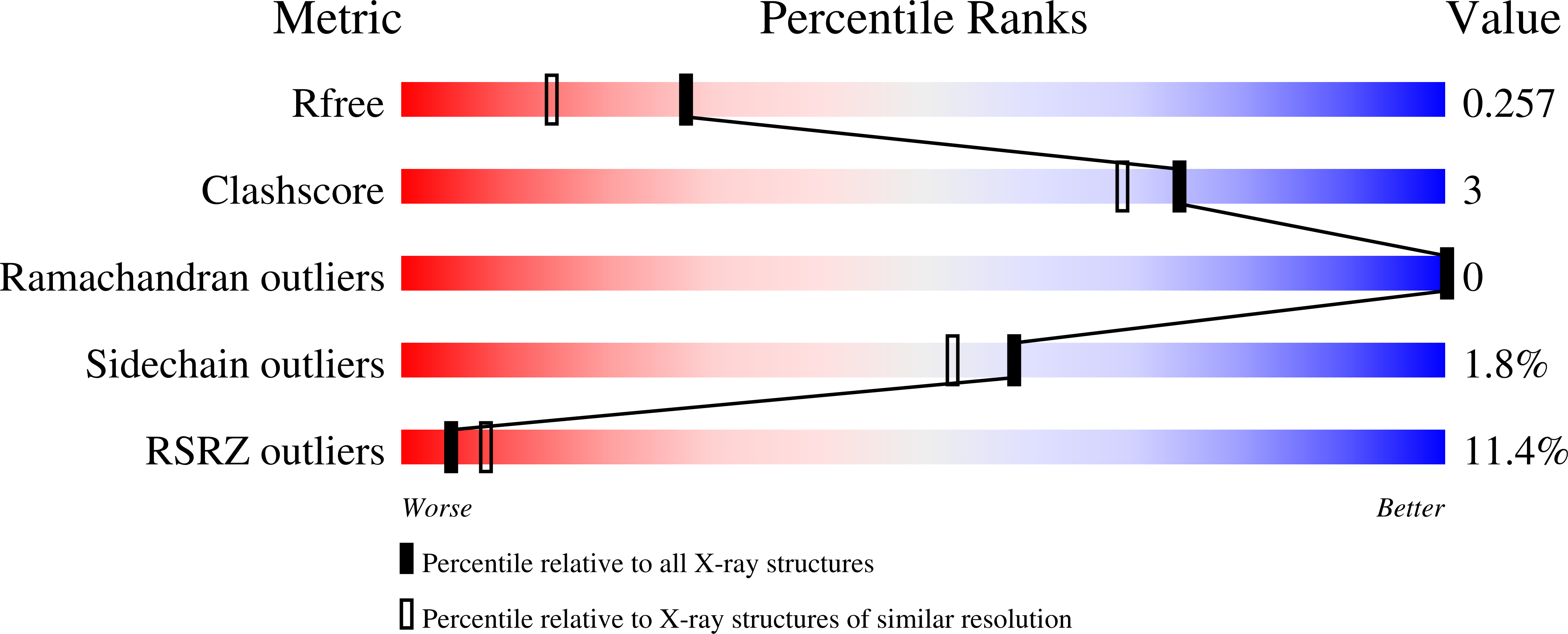
wwPDB Validation 3D Report Full Report
Entity ID: 1 | |||||
|---|---|---|---|---|---|
| Molecule | Chains | Sequence Length | Organism | Details | Image |
| Phosphomannomutase | 252 | Candida albicans SC5314 | Mutation(s): 0 Gene Names: PMM1, CaO19.10454, CaO19.2937 EC: 5.4.2.8 | 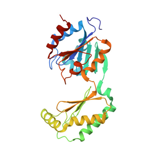 | |
UniProt | |||||
Find proteins for P31353 (Candida albicans (strain SC5314 / ATCC MYA-2876)) Explore P31353 Go to UniProtKB: P31353 | |||||
Entity Groups | |||||
| Sequence Clusters | 30% Identity50% Identity70% Identity90% Identity95% Identity100% Identity | ||||
| UniProt Group | P31353 | ||||
Sequence AnnotationsExpand | |||||
| |||||
| Ligands 2 Unique | |||||
|---|---|---|---|---|---|
| ID | Chains | Name / Formula / InChI Key | 2D Diagram | 3D Interactions | |
| CL Query on CL | E [auth A] | CHLORIDE ION Cl VEXZGXHMUGYJMC-UHFFFAOYSA-M |  | ||
| MG Query on MG | C [auth A], D [auth A], F [auth B], G [auth B] | MAGNESIUM ION Mg JLVVSXFLKOJNIY-UHFFFAOYSA-N |  | ||
| Length ( Å ) | Angle ( ˚ ) |
|---|---|
| a = 62.225 | α = 90 |
| b = 80.719 | β = 90 |
| c = 116.729 | γ = 90 |
| Software Name | Purpose |
|---|---|
| PHENIX | refinement |
| HKL-3000 | data reduction |
| HKL-3000 | data scaling |
| BALBES | phasing |
| PHENIX | model building |
| Coot | model building |
| Funding Organization | Location | Grant Number |
|---|---|---|
| National Institutes of Health/National Institute Of Allergy and Infectious Diseases (NIH/NIAID) | United States | HHSN272201200026C |
RCSB PDB (citation) is hosted by
RCSB PDB is a member of the

