5UAM

Structure of a new family of Polysaccharide lyase PL25-Ulvanlyase.


Experimental Data Snapshot

  • Method: X-RAY DIFFRACTION
  • Resolution: 1.45 Å
  • R-Value Free: 0.143 
  • R-Value Work: 0.134 
  • R-Value Observed: 0.134 

wwPDB Validation   3D Report Full Report


This is version 1.2 of the entry. See complete history


Literature

New Ulvan-Degrading Polysaccharide Lyase Family: Structure and Catalytic Mechanism Suggests Convergent Evolution of Active Site Architecture.

Ulaganathan, T.Boniecki, M.T.Foran, E.Buravenkov, V.Mizrachi, N.Banin, E.Helbert, W.Cygler, M.

(2017) ACS Chem Biol 12: 1269-1280

  • DOI: https://doi.org/10.1021/acschembio.7b00126
  • Primary Citation of Related Structures:  
    5UAM, 5UAS

  • PubMed Abstract: 

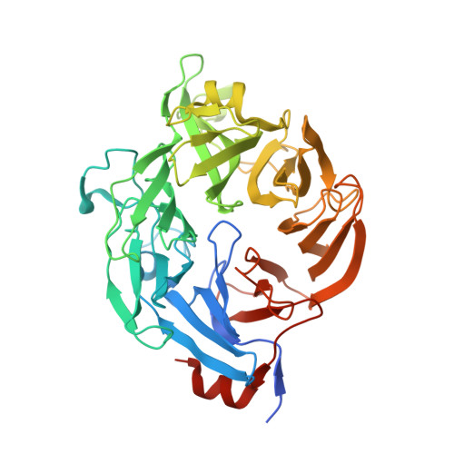
    Ulvan is a complex sulfated polysaccharide biosynthesized by green seaweed and contains predominantly rhamnose, xylose, and uronic acid sugars. Ulvan-degrading enzymes have only recently been identified and added to the CAZy ( www.cazy.org ) database as family PL24, but neither their structure nor catalytic mechanism(s) are yet known. Several homologous, new ulvan lyases, have been discovered in Pseudoalteromonas sp. strain PLSV, Alteromonas LOR, and Nonlabens ulvanivorans, defining a new family PL25, with the lyase encoded by the gene PLSV_3936 being one of them. This enzyme cleaves the glycosidic bond between 3-sulfated rhamnose (R3S) and glucuronic acid (GlcA) or iduronic acid (IdoA) via a β-elimination mechanism. We report the crystal structure of PLSV_3936 and its complex with a tetrasaccharide substrate. PLSV_3936 folds into a seven-bladed β-propeller, with each blade consisting of four antiparallel β-strands. Sequence conservation analysis identified a highly conserved region lining at one end of a deep crevice on the protein surface. The putative active site was identified by mutagenesis and activity measurements. Crystal structure of the enzyme with a bound tetrasaccharide substrate confirmed the identity of base and acid residues and allowed determination of the catalytic mechanism and also the identification of residues neutralizing the uronic acid carboxylic group. The PLSV_3936 structure provides an example of a convergent evolution among polysaccharide lyases toward a common active site architecture embedded in distinct folds.


  • Organizational Affiliation

    Department of Biochemistry, University of Saskatchewan , Saskatoon, Saskatchewan S7N 5E5, Canada.


Macromolecules
Find similar proteins by:  (by identity cutoff)  |  3D Structure
Entity ID: 1
MoleculeChains Sequence LengthOrganismDetailsImage
Ulvan Lyase-PL25
A, B
473Pseudoalteromonas sp. PLSVMutation(s): 0 
UniProt
Find proteins for A0A1W2VMZ5 (Pseudoalteromonas sp. (strain PLSV))
Explore A0A1W2VMZ5 
Go to UniProtKB:  A0A1W2VMZ5
Entity Groups  
Sequence Clusters30% Identity50% Identity70% Identity90% Identity95% Identity100% Identity
UniProt GroupA0A1W2VMZ5
Sequence Annotations
Expand
  • Reference Sequence
Small Molecules
Ligands 6 Unique
IDChains Name / Formula / InChI Key2D Diagram3D Interactions
PO4
Query on PO4

Download Ideal Coordinates CCD File 
E [auth A],
P [auth B]
PHOSPHATE ION
O4 P
NBIIXXVUZAFLBC-UHFFFAOYSA-K
GOL
Query on GOL

Download Ideal Coordinates CCD File 
F [auth A]GLYCEROL
C3 H8 O3
PEDCQBHIVMGVHV-UHFFFAOYSA-N
ZN
Query on ZN

Download Ideal Coordinates CCD File 
D [auth A],
O [auth B]
ZINC ION
Zn
PTFCDOFLOPIGGS-UHFFFAOYSA-N
EDO
Query on EDO

Download Ideal Coordinates CCD File 
H [auth A]
I [auth A]
J [auth A]
K [auth A]
L [auth A]
H [auth A],
I [auth A],
J [auth A],
K [auth A],
L [auth A],
M [auth A],
R [auth B],
S [auth B],
T [auth B]
1,2-ETHANEDIOL
C2 H6 O2
LYCAIKOWRPUZTN-UHFFFAOYSA-N
K
Query on K

Download Ideal Coordinates CCD File 
G [auth A],
Q [auth B]
POTASSIUM ION
K
NPYPAHLBTDXSSS-UHFFFAOYSA-N
CL
Query on CL

Download Ideal Coordinates CCD File 
C [auth A],
N [auth B]
CHLORIDE ION
Cl
VEXZGXHMUGYJMC-UHFFFAOYSA-M
Experimental Data & Validation

Experimental Data

  • Method: X-RAY DIFFRACTION
  • Resolution: 1.45 Å
  • R-Value Free: 0.143 
  • R-Value Work: 0.134 
  • R-Value Observed: 0.134 
  • Space Group: C 1 2 1
Unit Cell:
Length ( Å )Angle ( ˚ )
a = 174.26α = 90
b = 72.11β = 122.5
c = 109.51γ = 90
Software Package:
Software NamePurpose
PHENIXrefinement
XDSdata scaling
AutoSolphasing

Structure Validation

View Full Validation Report


Entry History 

Deposition Data

Revision History  (Full details and data files)

  • Version 1.0: 2017-03-29
    Type: Initial release
  • Version 1.1: 2017-05-31
    Changes: Database references
  • Version 1.2: 2024-03-06
    Changes: Data collection, Database references, Derived calculations