5TPB

Binding domain of BoNT/A complexed with ganglioside variant


Experimental Data Snapshot

  • Method: X-RAY DIFFRACTION
  • Resolution: 2.60 Å
  • R-Value Free: 0.240 
  • R-Value Work: 0.222 
  • R-Value Observed: 0.223 

wwPDB Validation   3D Report Full Report


Ligand Structure Quality Assessment 


This is version 2.0 of the entry. See complete history


Literature

Glycans Confer Specificity to the Recognition of Ganglioside Receptors by Botulinum Neurotoxin A.

Hamark, C.Berntsson, R.P.Masuyer, G.Henriksson, L.M.Gustafsson, R.Stenmark, P.Widmalm, G.

(2017) J Am Chem Soc 139: 218-230

  • DOI: https://doi.org/10.1021/jacs.6b09534
  • Primary Citation of Related Structures:  
    5TPB, 5TPC

  • PubMed Abstract: 

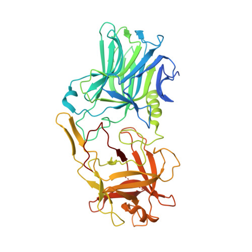
    The highly poisonous botulinum neurotoxins, produced by the bacterium Clostridium botulinum, act on their hosts by a high-affinity association to two receptors on neuronal cell surfaces as the first step of invasion. The glycan motifs of gangliosides serve as initial coreceptors for these protein complexes, whereby a membrane protein receptor is bound. Herein we set out to characterize the carbohydrate minimal binding epitope of the botulinum neurotoxin serotype A. By means of ligand-based NMR spectroscopy, X-ray crystallography, computer simulations, and isothermal titration calorimetry, a screening of ganglioside analogues together with a detailed characterization of various carbohydrate ligand complexes with the toxin were accomplished. We show that the representation of the glycan epitope to the protein affects the details of binding. Notably, both branches of the oligosaccharide GD1a can associate to botulinum neurotoxin serotype A when expressed as individual trisaccharides. It is, however, the terminal branch of GD1a as well as this trisaccharide motif alone, corresponding to the sialyl-Thomsen-Friedenreich antigen, that represents the active ligand epitope, and these compounds bind to the neurotoxin with a high degree of predisposition but with low affinities. This finding does not correlate with the oligosaccharide moieties having a strong contribution to the total affinity, which was expected to be the case. We here propose that the glycan part of the ganglioside receptors mainly provides abundance and specificity, whereas the interaction with the membrane itself and protein receptor brings about the strong total binding of the toxin to the neuronal membrane.


  • Organizational Affiliation

    Department of Organic Chemistry, Arrhenius Laboratory, Stockholm University , S-106 91 Stockholm, Sweden.


Macromolecules
Find similar proteins by:  (by identity cutoff)  |  3D Structure
Entity ID: 1
MoleculeChains Sequence LengthOrganismDetailsImage
Botulinum neurotoxin type A
A, B
444Clostridium botulinumMutation(s): 0 
Gene Names: botAatxbna
EC: 3.4.24.69
UniProt
Find proteins for P0DPI1 (Clostridium botulinum (strain Hall / ATCC 3502 / NCTC 13319 / Type A))
Explore P0DPI1 
Go to UniProtKB:  P0DPI1
Entity Groups  
Sequence Clusters30% Identity50% Identity70% Identity90% Identity95% Identity100% Identity
UniProt GroupP0DPI1
Sequence Annotations
Expand
  • Reference Sequence
Oligosaccharides

Help

Entity ID: 2
MoleculeChains Length2D Diagram Glycosylation3D Interactions
beta-D-galactopyranose-(1-3)-2-acetamido-2-deoxy-beta-D-galactopyranose
C
2N/A
Glycosylation Resources
GlyTouCan:  G01534TU
GlyCosmos:  G01534TU
GlyGen:  G01534TU
Small Molecules
Ligands 1 Unique
IDChains Name / Formula / InChI Key2D Diagram3D Interactions
SIA
Query on SIA

Download Ideal Coordinates CCD File 
D [auth B]N-acetyl-alpha-neuraminic acid
C11 H19 N O9
SQVRNKJHWKZAKO-YRMXFSIDSA-N
Experimental Data & Validation

Experimental Data

  • Method: X-RAY DIFFRACTION
  • Resolution: 2.60 Å
  • R-Value Free: 0.240 
  • R-Value Work: 0.222 
  • R-Value Observed: 0.223 
  • Space Group: P 1 21 1
Unit Cell:
Length ( Å )Angle ( ˚ )
a = 65.515α = 90
b = 104.33β = 115.97
c = 69.406γ = 90
Software Package:
Software NamePurpose
REFMACrefinement
SCALAdata scaling
PHASERphasing
PDB_EXTRACTdata extraction

Structure Validation

View Full Validation Report


Ligand Structure Quality Assessment 


Entry History 

Deposition Data

Revision History  (Full details and data files)

  • Version 1.0: 2017-01-18
    Type: Initial release
  • Version 2.0: 2020-07-29
    Type: Remediation
    Reason: Carbohydrate remediation
    Changes: Advisory, Atomic model, Data collection, Derived calculations, Structure summary