5T4D

Cryo-EM structure of Polycystic Kidney Disease protein 2 (PKD2), residues 198-703


Experimental Data Snapshot

  • Method: ELECTRON MICROSCOPY
  • Resolution: 3.00 Å
  • Aggregation State: PARTICLE 
  • Reconstruction Method: SINGLE PARTICLE 

wwPDB Validation   3D Report Full Report


This is version 1.5 of the entry. See complete history


Literature

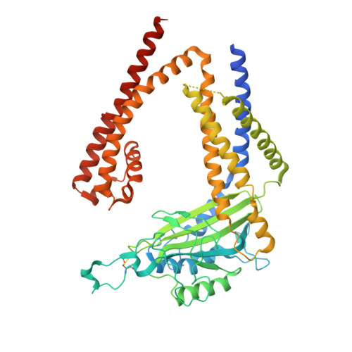
The Structure of the Polycystic Kidney Disease Channel PKD2 in Lipid Nanodiscs.

Shen, P.S.Yang, X.DeCaen, P.G.Liu, X.Bulkley, D.Clapham, D.E.Cao, E.

(2016) Cell 167: 763-773.e11

  • DOI: https://doi.org/10.1016/j.cell.2016.09.048
  • Primary Citation of Related Structures:  
    5T4D

  • PubMed Abstract: 

    The Polycystic Kidney Disease 2 (Pkd2) gene is mutated in autosomal dominant polycystic kidney disease (ADPKD), one of the most common human monogenic disorders. Here, we present the cryo-EM structure of PKD2 in lipid bilayers at 3.0 Å resolution, which establishes PKD2 as a homotetrameric ion channel and provides insight into potential mechanisms for its activation. The PKD2 voltage-sensor domain retains two of four gating charges commonly found in those of voltage-gated ion channels. The PKD2 ion permeation pathway is constricted at the selectivity filter and near the cytoplasmic end of S6, suggesting that two gates regulate ion conduction. The extracellular domain of PKD2, a hotspot for ADPKD pathogenic mutations, contributes to channel assembly and strategically interacts with the transmembrane core, likely serving as a physical substrate for extracellular stimuli to allosterically gate the channel. Finally, our structure establishes the molecular basis for the majority of pathogenic mutations in Pkd2-related ADPKD.


  • Organizational Affiliation

    Department of Biochemistry, University of Utah School of Medicine, Salt Lake City, UT 84112-5650, USA.


Macromolecules
Find similar proteins by:  (by identity cutoff)  |  3D Structure
Entity ID: 1
MoleculeChains Sequence LengthOrganismDetailsImage
hPKD:198-703, Polycystin-2
A, B, C, D
510Homo sapiensMutation(s): 0 
Gene Names: PKD2TRPP2
Membrane Entity: Yes 
UniProt & NIH Common Fund Data Resources
Find proteins for Q13563 (Homo sapiens)
Explore Q13563 
Go to UniProtKB:  Q13563
PHAROS:  Q13563
GTEx:  ENSG00000118762 
Entity Groups  
Sequence Clusters30% Identity50% Identity70% Identity90% Identity95% Identity100% Identity
UniProt GroupQ13563
Sequence Annotations
Expand
  • Reference Sequence
Small Molecules
Ligands 1 Unique
IDChains Name / Formula / InChI Key2D Diagram3D Interactions
NAG
Query on NAG

Download Ideal Coordinates CCD File 
E [auth A]
F [auth A]
G [auth A]
H [auth B]
I [auth B]
E [auth A],
F [auth A],
G [auth A],
H [auth B],
I [auth B],
J [auth B],
K [auth C],
L [auth C],
M [auth C],
N [auth D],
O [auth D],
P [auth D]
2-acetamido-2-deoxy-beta-D-glucopyranose
C8 H15 N O6
OVRNDRQMDRJTHS-FMDGEEDCSA-N
Experimental Data & Validation

Experimental Data

  • Method: ELECTRON MICROSCOPY
  • Resolution: 3.00 Å
  • Aggregation State: PARTICLE 
  • Reconstruction Method: SINGLE PARTICLE 
EM Software:
TaskSoftware PackageVersion
RECONSTRUCTIONRELION1.4
MODEL REFINEMENTPHENIX1.10.1-2155

Structure Validation

View Full Validation Report


Entry History 

Deposition Data

Revision History  (Full details and data files)

  • Version 1.0: 2016-11-02
    Type: Initial release
  • Version 1.1: 2016-11-09
    Changes: Structure summary
  • Version 1.2: 2016-11-30
    Changes: Structure summary
  • Version 1.3: 2017-11-08
    Changes: Data processing, Derived calculations
  • Version 1.4: 2019-11-06
    Changes: Data collection, Other
  • Version 1.5: 2020-07-29
    Type: Remediation
    Reason: Carbohydrate remediation
    Changes: Data collection, Derived calculations, Structure summary