5SVW

Light-state Structure of Arabidopsis Thaliana Zeitlupe


Experimental Data Snapshot

  • Method: X-RAY DIFFRACTION
  • Resolution: 2.29 Å
  • R-Value Free: 0.228 
  • R-Value Work: 0.166 
  • R-Value Observed: 0.169 

wwPDB Validation   3D Report Full Report


Ligand Structure Quality Assessment 


This is version 1.6 of the entry. See complete history


Literature

Kinetics of the LOV domain of ZEITLUPE determine its circadian function inArabidopsis.

Pudasaini, A.Shim, J.S.Song, Y.H.Shi, H.Kiba, T.Somers, D.E.Imaizumi, T.Zoltowski, B.D.

(2017) Elife 6

  • DOI: https://doi.org/10.7554/eLife.21646
  • Primary Citation of Related Structures:  
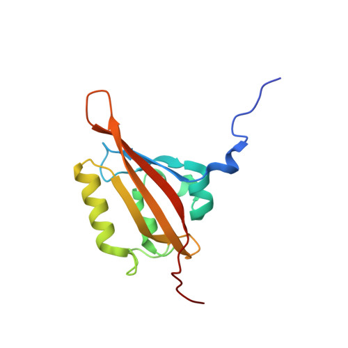
    5SVG, 5SVU, 5SVV, 5SVW

  • PubMed Abstract: 

    A LOV (Light, Oxygen, or Voltage) domain containing blue-light photoreceptor ZEITLUPE (ZTL) directs circadian timing by degrading clock proteins in plants. Functions hinge upon allosteric differences coupled to the ZTL photocycle; however, structural and kinetic information was unavailable. Herein, we tune the ZTL photocycle over two orders of magnitude. These variants reveal that ZTL complexes with targets independent of light, but dictates enhanced protein degradation in the dark. In vivo experiments definitively show photocycle kinetics dictate the rate of clock component degradation, thereby impacting circadian period. Structural studies demonstrate that photocycle dependent activation of ZTL depends on an unusual dark-state conformation of ZTL. Crystal structures of ZTL LOV domain confirm delineation of structural and kinetic mechanisms and identify an evolutionarily selected allosteric hinge differentiating modes of PAS/LOV signal transduction. The combined biochemical, genetic and structural studies provide new mechanisms indicating how PAS/LOV proteins integrate environmental variables in complex networks.


  • Organizational Affiliation

    Department of Chemistry, Southern Methodist University, Dallas, United States.


Macromolecules
Find similar proteins by:  (by identity cutoff)  |  3D Structure
Entity ID: 1
MoleculeChains Sequence LengthOrganismDetailsImage
Adagio protein 1A [auth B],
B [auth A],
C,
D
137Arabidopsis thalianaMutation(s): 2 
Gene Names: ADO1FKL2LKP1ZTLAt5g57360MSF19.2
UniProt
Find proteins for Q94BT6 (Arabidopsis thaliana)
Explore Q94BT6 
Go to UniProtKB:  Q94BT6
Entity Groups  
Sequence Clusters30% Identity50% Identity70% Identity90% Identity95% Identity100% Identity
UniProt GroupQ94BT6
Sequence Annotations
Expand
  • Reference Sequence
Experimental Data & Validation

Experimental Data

  • Method: X-RAY DIFFRACTION
  • Resolution: 2.29 Å
  • R-Value Free: 0.228 
  • R-Value Work: 0.166 
  • R-Value Observed: 0.169 
  • Space Group: P 31 2 1
Unit Cell:
Length ( Å )Angle ( ˚ )
a = 86.221α = 90
b = 86.221β = 90
c = 200.478γ = 120
Software Package:
Software NamePurpose
PHENIXrefinement
HKL-2000data reduction
HKL-2000data scaling
PHENIXphasing

Structure Validation

View Full Validation Report


Ligand Structure Quality Assessment 


Entry History & Funding Information

Deposition Data


Funding OrganizationLocationGrant Number
National Institutes of Health/National Institute of General Medical Sciences (NIH/NIGMS)United StatesR15GM109282

Revision History  (Full details and data files)

  • Version 1.0: 2017-03-08
    Type: Initial release
  • Version 1.1: 2017-03-15
    Changes: Database references
  • Version 1.2: 2017-09-27
    Changes: Author supporting evidence
  • Version 1.3: 2018-02-28
    Changes: Database references
  • Version 1.4: 2018-03-28
    Changes: Data collection, Database references
  • Version 1.5: 2019-12-25
    Changes: Author supporting evidence, Derived calculations
  • Version 1.6: 2023-10-04
    Changes: Data collection, Database references, Derived calculations, Refinement description