High-Throughput Crystallography: Reliable and Efficient Identification of Fragment Hits.
Schiebel, J., Krimmer, S.G., Rower, K., Knorlein, A., Wang, X., Park, A.Y., Stieler, M., Ehrmann, F.R., Fu, K., Radeva, N., Krug, M., Huschmann, F.U., Glockner, S., Weiss, M.S., Mueller, U., Klebe, G., Heine, A.(2016) Structure 24: 1398-1409
- PubMed: 27452405
- DOI: https://doi.org/10.1016/j.str.2016.06.010
- Primary Citation of Related Structures:
5OYQ, 5OYR, 5OYS, 5OYT, 5OYU, 5OYV, 5OYW, 5OYX, 5OYY, 5OYZ, 5OZ0, 5OZ1, 5OZ2, 5OZ3, 5OZ4, 5OZ5, 5OZ6, 5OZ7, 5OZ8, 5OZ9, 5OZA, 5OZB, 5OZC, 5OZD, 5OZE, 5OZF, 5OZG, 5OZH, 5OZI, 5OZJ, 5OZK, 5OZL, 5OZM, 5OZN, 5OZO, 5OZP, 5OZQ, 5OZR, 5OZS, 5OZT, 5OZU, 5OZV, 5OZW, 5OZX, 5OZY, 5OZZ, 5P00, 5P01, 5P02, 5P03 - PubMed Abstract:
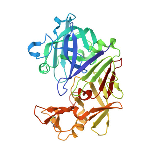
Today the identification of lead structures for drug development often starts from small fragment-like molecules raising the chances to find compounds that successfully pass clinical trials. At the heart of the screening for fragments binding to a specific target, crystallography delivers structural information essential for subsequent drug design. While it is common to search for bound ligands in electron densities calculated directly after an initial refinement cycle, we raise the important question whether this strategy is viable for fragments characterized by low affinities. Here, we describe and provide a collection of high-quality diffraction data obtained from 364 protein crystals treated with diverse fragments. Subsequent data analysis showed that ∼25% of all hits would have been missed without further refining the resulting structures. To enable fast and reliable hit identification, we have designed an automated refinement pipeline that will inspire the development of optimized tools facilitating the successful application of fragment-based methods.
Organizational Affiliation:
Institute for Pharmaceutical Chemistry, Philipps-University Marburg, Marbacher Weg 6, 35032 Marburg, Germany.