5OYH

crystal structure of the catalytic core of a rhodopsin-guanylyl cyclase with converted specificity in complex with ATPalphaS


Experimental Data Snapshot

  • Method: X-RAY DIFFRACTION
  • Resolution: 2.25 Å
  • R-Value Free: 0.224 
  • R-Value Work: 0.182 
  • R-Value Observed: 0.183 

wwPDB Validation   3D Report Full Report


This is version 1.0 of the entry. See complete history


Literature

Rhodopsin-cyclases for photocontrol of cGMP/cAMP and 2.3 angstrom structure of the adenylyl cyclase domain.

Scheib, U.Broser, M.Constantin, O.M.Yang, S.Gao, S.Mukherjee, S.Stehfest, K.Nagel, G.Gee, C.E.Hegemann, P.

(2018) Nat Commun 9: 2046-2046

  • DOI: https://doi.org/10.1038/s41467-018-04428-w
  • Primary Citation of Related Structures:  
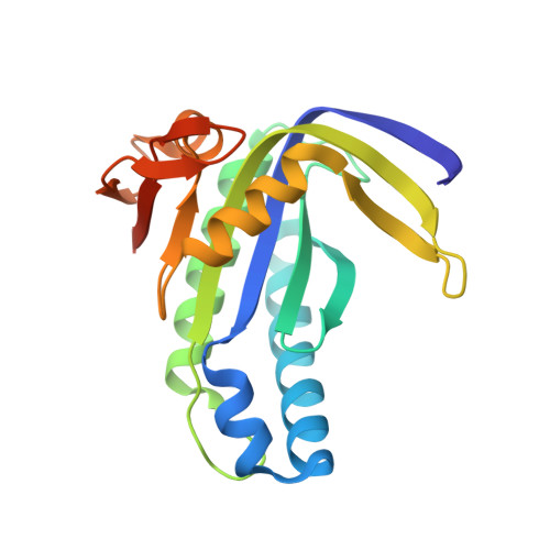
    5OYH

  • PubMed Abstract: 

    The cyclic nucleotides cAMP and cGMP are important second messengers that orchestrate fundamental cellular responses. Here, we present the characterization of the rhodopsin-guanylyl cyclase from Catenaria anguillulae (CaRhGC), which produces cGMP in response to green light with a light to dark activity ratio >1000. After light excitation the putative signaling state forms with τ = 31 ms and decays with τ = 570 ms. Mutations (up to 6) within the nucleotide binding site generate rhodopsin-adenylyl cyclases (CaRhACs) of which the double mutated YFP-CaRhAC (E497K/C566D) is the most suitable for rapid cAMP production in neurons. Furthermore, the crystal structure of the ligand-bound AC domain (2.25 Å) reveals detailed information about the nucleotide binding mode within this recently discovered class of enzyme rhodopsin. Both YFP-CaRhGC and YFP-CaRhAC are favorable optogenetic tools for non-invasive, cell-selective, and spatio-temporally precise modulation of cAMP/cGMP with light.


  • Organizational Affiliation

    Institute for Biology, Experimental Biophysics, Humboldt-Universität zu Berlin, 10115, Berlin, Germany.


Macromolecules
Find similar proteins by:  (by identity cutoff)  |  3D Structure
Entity ID: 1
MoleculeChains Sequence LengthOrganismDetailsImage
Nucleotide cyclase
A, B, C, D, E
A, B, C, D, E, F, G, H, I, J, K, L, M, N, O, P
193Catenaria anguillulae PL171Mutation(s): 2 
Gene Names: BCR44DRAFT_1480233
UniProt
Find proteins for A0A1Y2HEJ3 (Catenaria anguillulae PL171)
Explore A0A1Y2HEJ3 
Go to UniProtKB:  A0A1Y2HEJ3
Entity Groups  
Sequence Clusters30% Identity50% Identity70% Identity90% Identity95% Identity100% Identity
UniProt GroupA0A1Y2HEJ3
Sequence Annotations
Expand
  • Reference Sequence
Small Molecules
Ligands 3 Unique
IDChains Name / Formula / InChI Key2D Diagram3D Interactions
T99
Query on T99

Download Ideal Coordinates CCD File 
AA [auth D]
AB [auth L]
CB [auth M]
DA [auth E]
EB [auth N]
AA [auth D],
AB [auth L],
CB [auth M],
DA [auth E],
EB [auth N],
GB [auth O],
HA [auth F],
JB [auth P],
KA [auth G],
NA [auth H],
Q [auth A],
QA [auth I],
U [auth B],
UA [auth J],
WA [auth K],
X [auth C]
ADENOSINE-5'-SP-ALPHA-THIO-TRIPHOSPHATE
C10 H16 N5 O12 P3 S
ROYJKVPBJVNHCQ-CENJAJHPSA-N
GOL
Query on GOL

Download Ideal Coordinates CCD File 
BA [auth D]
FA [auth E]
GA [auth F]
IA [auth F]
IB [auth O]
BA [auth D],
FA [auth E],
GA [auth F],
IA [auth F],
IB [auth O],
MA [auth G],
PA [auth I],
S [auth A],
SA [auth J],
T [auth A],
TA [auth J],
W [auth C],
YA [auth K],
Z [auth C],
ZA [auth L]
GLYCEROL
C3 H8 O3
PEDCQBHIVMGVHV-UHFFFAOYSA-N
CA
Query on CA

Download Ideal Coordinates CCD File 
BB [auth L]
CA [auth D]
DB [auth M]
EA [auth E]
FB [auth N]
BB [auth L],
CA [auth D],
DB [auth M],
EA [auth E],
FB [auth N],
HB [auth O],
JA [auth F],
KB [auth P],
LA [auth G],
OA [auth H],
R [auth A],
RA [auth I],
V [auth B],
VA [auth J],
XA [auth K],
Y [auth C]
CALCIUM ION
Ca
BHPQYMZQTOCNFJ-UHFFFAOYSA-N
Experimental Data & Validation

Experimental Data

  • Method: X-RAY DIFFRACTION
  • Resolution: 2.25 Å
  • R-Value Free: 0.224 
  • R-Value Work: 0.182 
  • R-Value Observed: 0.183 
  • Space Group: I 41
Unit Cell:
Length ( Å )Angle ( ˚ )
a = 193.283α = 90
b = 193.283β = 90
c = 225.501γ = 90
Software Package:
Software NamePurpose
PHENIXrefinement
XDSdata reduction
PDB_EXTRACTdata extraction
PHENIXphasing
Cootmodel building

Structure Validation

View Full Validation Report


Entry History 

Deposition Data

Revision History  (Full details and data files)

  • Version 1.0: 2018-06-06
    Type: Initial release