5OUI

Humanized alpha-AChBP (acetylcholine binding protein) in complex with allosteric binder fragment CU2017


Experimental Data Snapshot

  • Method: X-RAY DIFFRACTION
  • Resolution: 3.10 Å
  • R-Value Free: 0.280 
  • R-Value Work: 0.241 
  • R-Value Observed: 0.243 

wwPDB Validation   3D Report Full Report


Ligand Structure Quality Assessment 


This is version 2.1 of the entry. See complete history


Literature

An allosteric binding site of the alpha 7 nicotinic acetylcholine receptor revealed in a humanized acetylcholine-binding protein.

Delbart, F.Brams, M.Gruss, F.Noppen, S.Peigneur, S.Boland, S.Chaltin, P.Brandao-Neto, J.von Delft, F.Touw, W.G.Joosten, R.P.Liekens, S.Tytgat, J.Ulens, C.

(2018) J Biol Chem 293: 2534-2545

  • DOI: https://doi.org/10.1074/jbc.M117.815316
  • Primary Citation of Related Structures:  
    5OUG, 5OUH, 5OUI

  • PubMed Abstract: 

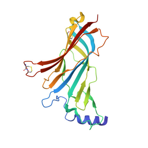
    Nicotinic acetylcholine receptors (nAChRs) belong to the family of pentameric ligand-gated ion channels and mediate fast excitatory transmission in the central and peripheral nervous systems. Among the different existing receptor subtypes, the homomeric α7 nAChR has attracted considerable attention because of its possible implication in several neurological and psychiatric disorders, including cognitive decline associated with Alzheimer's disease or schizophrenia. Allosteric modulators of ligand-gated ion channels are of particular interest as therapeutic agents, as they modulate receptor activity without affecting normal fluctuations of synaptic neurotransmitter release. Here, we used X-ray crystallography and surface plasmon resonance spectroscopy of α7-acetylcholine-binding protein (AChBP), a humanized chimera of a snail AChBP, which has 71% sequence similarity with the extracellular ligand-binding domain of the human α7 nAChR, to investigate the structural determinants of allosteric modulation. We extended previous observations that an allosteric site located in the vestibule of the receptor offers an attractive target for receptor modulation. We introduced seven additional humanizing mutations in the vestibule-located binding site of AChBP to improve its suitability as a model for studying allosteric binding. Using a fragment-based screening approach, we uncovered an allosteric binding site located near the β8-β9 loop, which critically contributes to coupling ligand binding to channel opening in human α7 nAChR. This work expands our understanding of the topology of allosteric binding sites in AChBP and, by extrapolation, in the human α7 nAChR as determined by electrophysiology measurements. Our insights pave the way for drug design strategies targeting nAChRs involved in ion channel-mediated disorders.


  • Organizational Affiliation

    From the Department of Cellular and Molecular Medicine, Laboratory of Structural Neurobiology, Faculty of Medicine, KU Leuven, 3000 Leuven, Belgium.


Macromolecules
Find similar proteins by:  (by identity cutoff)  |  3D Structure
Entity ID: 1
MoleculeChains Sequence LengthOrganismDetailsImage
Acetylcholine binding protein
A, B, C, D, E
205Homo sapiensLymnaea stagnalis
This entity is chimeric
Mutation(s): 0 
Entity Groups  
Sequence Clusters30% Identity50% Identity70% Identity90% Identity95% Identity100% Identity
Sequence Annotations
Expand
  • Reference Sequence
Oligosaccharides

Help

Entity ID: 2
MoleculeChains Length2D Diagram Glycosylation3D Interactions
alpha-D-mannopyranose-(1-3)-[alpha-D-mannopyranose-(1-6)]beta-D-mannopyranose-(1-4)-2-acetamido-2-deoxy-beta-D-glucopyranose-(1-4)-2-acetamido-2-deoxy-beta-D-glucopyranose
F
5N-Glycosylation
Glycosylation Resources
GlyTouCan:  G22768VO
GlyCosmos:  G22768VO
GlyGen:  G22768VO
Entity ID: 3
MoleculeChains Length2D Diagram Glycosylation3D Interactions
2-acetamido-2-deoxy-beta-D-glucopyranose-(1-4)-2-acetamido-2-deoxy-beta-D-glucopyranose
G, I
2N-Glycosylation
Glycosylation Resources
GlyTouCan:  G42666HT
GlyCosmos:  G42666HT
GlyGen:  G42666HT
Entity ID: 4
MoleculeChains Length2D Diagram Glycosylation3D Interactions
beta-D-mannopyranose-(1-4)-2-acetamido-2-deoxy-beta-D-glucopyranose-(1-4)-2-acetamido-2-deoxy-beta-D-glucopyranose
H, J
3N-Glycosylation
Glycosylation Resources
GlyTouCan:  G15407YE
GlyCosmos:  G15407YE
GlyGen:  G15407YE
Small Molecules
Ligands 3 Unique
IDChains Name / Formula / InChI Key2D Diagram3D Interactions
AVB
Query on AVB

Download Ideal Coordinates CCD File 
K [auth A](3~{R})-~{N}-(5-bromanylpyridin-2-yl)piperidine-3-carboxamide
C11 H14 Br N3 O
LURZDHQDKHLCGG-MRVPVSSYSA-N
PEG
Query on PEG

Download Ideal Coordinates CCD File 
BA [auth C]DI(HYDROXYETHYL)ETHER
C4 H10 O3
MTHSVFCYNBDYFN-UHFFFAOYSA-N
EDO
Query on EDO

Download Ideal Coordinates CCD File 
AA [auth C]
CA [auth D]
DA [auth D]
EA [auth D]
FA [auth D]
AA [auth C],
CA [auth D],
DA [auth D],
EA [auth D],
FA [auth D],
GA [auth D],
HA [auth E],
IA [auth E],
JA [auth E],
KA [auth E],
L [auth A],
LA [auth E],
M [auth A],
N [auth A],
O [auth A],
P [auth A],
Q [auth A],
R [auth A],
S [auth A],
T [auth A],
U [auth B],
V [auth B],
W [auth B],
X [auth B],
Y [auth B],
Z [auth C]
1,2-ETHANEDIOL
C2 H6 O2
LYCAIKOWRPUZTN-UHFFFAOYSA-N
Experimental Data & Validation

Experimental Data

  • Method: X-RAY DIFFRACTION
  • Resolution: 3.10 Å
  • R-Value Free: 0.280 
  • R-Value Work: 0.241 
  • R-Value Observed: 0.243 
  • Space Group: P 21 21 21
Unit Cell:
Length ( Å )Angle ( ˚ )
a = 87.753α = 90
b = 113.139β = 90
c = 145.887γ = 90
Software Package:
Software NamePurpose
REFMACrefinement
PDB_EXTRACTdata extraction
SCALAdata scaling
DIMPLEphasing
XDSdata reduction
Aimlessdata scaling

Structure Validation

View Full Validation Report


Ligand Structure Quality Assessment 


Entry History 

Deposition Data

Revision History  (Full details and data files)

  • Version 1.0: 2017-11-29
    Type: Initial release
  • Version 1.1: 2017-12-20
    Changes: Database references
  • Version 1.2: 2017-12-27
    Changes: Database references
  • Version 1.3: 2018-02-28
    Changes: Database references
  • Version 1.4: 2019-10-16
    Changes: Data collection
  • Version 2.0: 2020-07-29
    Type: Remediation
    Reason: Carbohydrate remediation
    Changes: Atomic model, Data collection, Derived calculations, Structure summary
  • Version 2.1: 2024-01-17
    Changes: Data collection, Database references, Derived calculations, Refinement description, Structure summary