5OQD

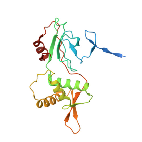
PHD2 and winged-helix domain of Polycomblike


Experimental Data Snapshot

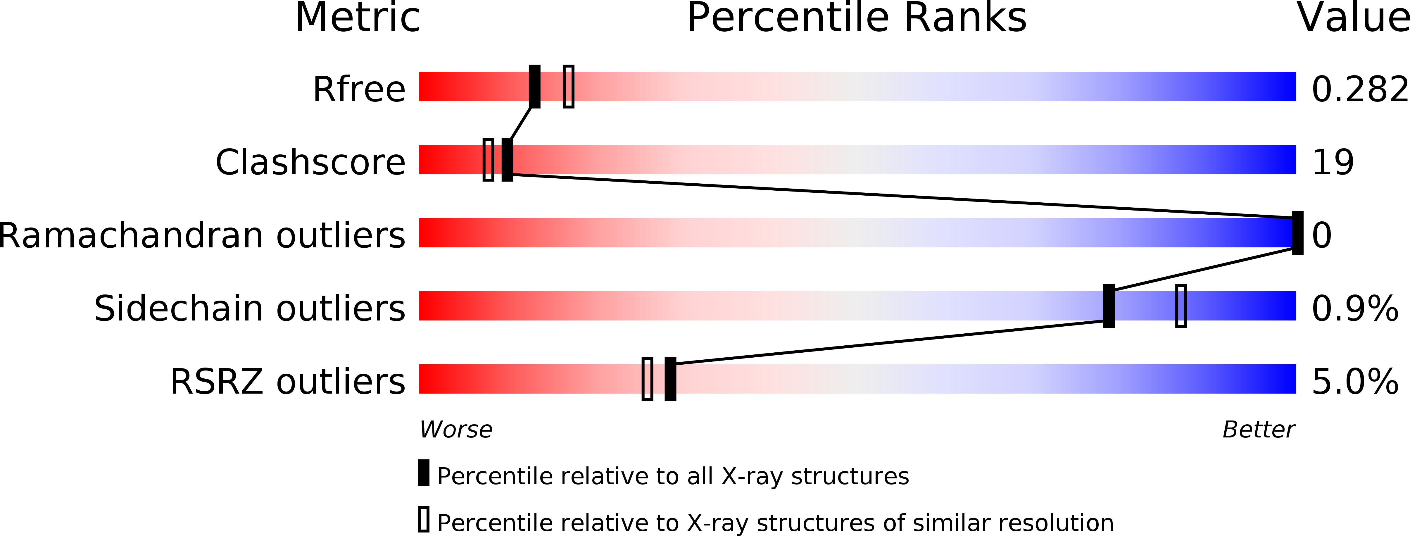
  • Method: X-RAY DIFFRACTION
  • Resolution: 2.45 Å
  • R-Value Free: 0.278 
  • R-Value Work: 0.236 
  • R-Value Observed: 0.238 

wwPDB Validation   3D Report Full Report


This is version 1.2 of the entry. See complete history


Literature

DNA binding by PHF1 prolongs PRC2 residence time on chromatin and thereby promotes H3K27 methylation.

Choi, J.Bachmann, A.L.Tauscher, K.Benda, C.Fierz, B.Muller, J.

(2017) Nat Struct Mol Biol 24: 1039-1047

  • DOI: https://doi.org/10.1038/nsmb.3488
  • Primary Citation of Related Structures:  
    5OQD

  • PubMed Abstract: 

    Polycomb repressive complex 2 (PRC2) trimethylates histone H3 at lysine 27 to mark genes for repression. We measured the dynamics of PRC2 binding on recombinant chromatin and free DNA at the single-molecule level using total internal reflection fluorescence (TIRF) microscopy. PRC2 preferentially binds free DNA with multisecond residence time and midnanomolar affinity. PHF1, a PRC2 accessory protein of the Polycomblike family, extends PRC2 residence time on DNA and chromatin. Crystallographic and functional studies reveal that Polycomblike proteins contain a winged-helix domain that binds DNA in a sequence-nonspecific fashion. DNA binding by this winged-helix domain accounts for the prolonged residence time of PHF1-PRC2 on chromatin and makes it a more efficient H3K27 methyltranferase than PRC2 alone. Together, these studies establish that interactions with DNA provide the predominant binding affinity of PRC2 for chromatin. Moreover, they reveal the molecular basis for how Polycomblike proteins stabilize PRC2 on chromatin and stimulate its activity.


  • Organizational Affiliation

    Max Planck Institute of Biochemistry, Laboratory of Chromatin Biology, Martinsried, Germany.


Macromolecules
Find similar proteins by:  (by identity cutoff)  |  3D Structure
Entity ID: 1
MoleculeChains Sequence LengthOrganismDetailsImage
Polycomb protein Pcl209Drosophila melanogasterMutation(s): 0 
Gene Names: PclCG5109
UniProt
Find proteins for Q24459 (Drosophila melanogaster)
Explore Q24459 
Go to UniProtKB:  Q24459
Entity Groups  
Sequence Clusters30% Identity50% Identity70% Identity90% Identity95% Identity100% Identity
UniProt GroupQ24459
Sequence Annotations
Expand
  • Reference Sequence
Small Molecules
Ligands 3 Unique
IDChains Name / Formula / InChI Key2D Diagram3D Interactions
PO4
Query on PO4

Download Ideal Coordinates CCD File 
O [auth C],
T [auth D]
PHOSPHATE ION
O4 P
NBIIXXVUZAFLBC-UHFFFAOYSA-K
ZN
Query on ZN

Download Ideal Coordinates CCD File 
G [auth B]
H [auth B]
K [auth A]
L [auth A]
M [auth C]
G [auth B],
H [auth B],
K [auth A],
L [auth A],
M [auth C],
N [auth C],
R [auth D],
S [auth D],
U [auth E],
V [auth E],
W [auth F],
X [auth F]
ZINC ION
Zn
PTFCDOFLOPIGGS-UHFFFAOYSA-N
EDO
Query on EDO

Download Ideal Coordinates CCD File 
I [auth B],
J [auth B],
P [auth C],
Q [auth C],
Y [auth F]
1,2-ETHANEDIOL
C2 H6 O2
LYCAIKOWRPUZTN-UHFFFAOYSA-N
Experimental Data & Validation

Experimental Data

  • Method: X-RAY DIFFRACTION
  • Resolution: 2.45 Å
  • R-Value Free: 0.278 
  • R-Value Work: 0.236 
  • R-Value Observed: 0.238 
  • Space Group: C 1 2 1
Unit Cell:
Length ( Å )Angle ( ˚ )
a = 307.423α = 90
b = 53.119β = 105.47
c = 86.839γ = 90
Software Package:
Software NamePurpose
PHENIXrefinement
XDSdata reduction
XDSdata scaling
PHENIXphasing

Structure Validation

View Full Validation Report


Entry History & Funding Information

Deposition Data


Funding OrganizationLocationGrant Number
European CommissionGermany277899

Revision History  (Full details and data files)

  • Version 1.0: 2017-11-01
    Type: Initial release
  • Version 1.1: 2017-12-20
    Changes: Database references
  • Version 1.2: 2024-05-08
    Changes: Data collection, Database references