5OLN

X-Ray Structure of the Complex Pyrimidine-nucleoside phosphorylase from Bacillus subtilis at 1.88 A


Experimental Data Snapshot

  • Method: X-RAY DIFFRACTION
  • Resolution: 1.88 Å
  • R-Value Free: 0.239 
  • R-Value Work: 0.200 
  • R-Value Observed: 0.201 

wwPDB Validation   3D Report Full Report


This is version 1.2 of the entry. See complete history


Literature

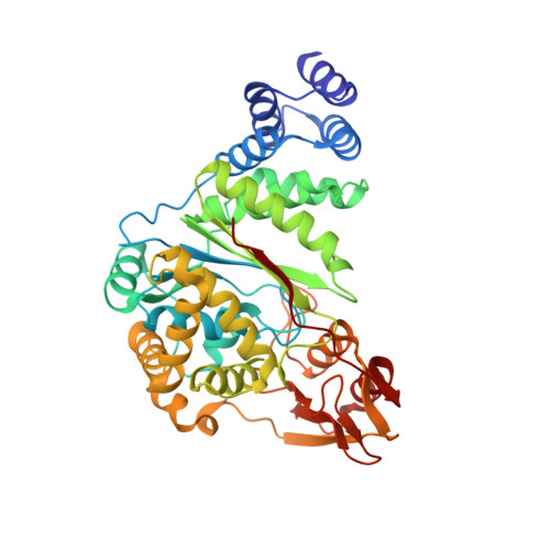
Crystal structure of pyrimidine-nucleoside phosphorylase from Bacillus subtilis in complex with imidazole and sulfate.

Balaev, V.V.Prokofev, I.I.Gabdoulkhakov, A.G.Betzel, C.Lashkov, A.A.

(2018) Acta Crystallogr F Struct Biol Commun 74: 193-197

  • DOI: https://doi.org/10.1107/S2053230X18002935
  • Primary Citation of Related Structures:  
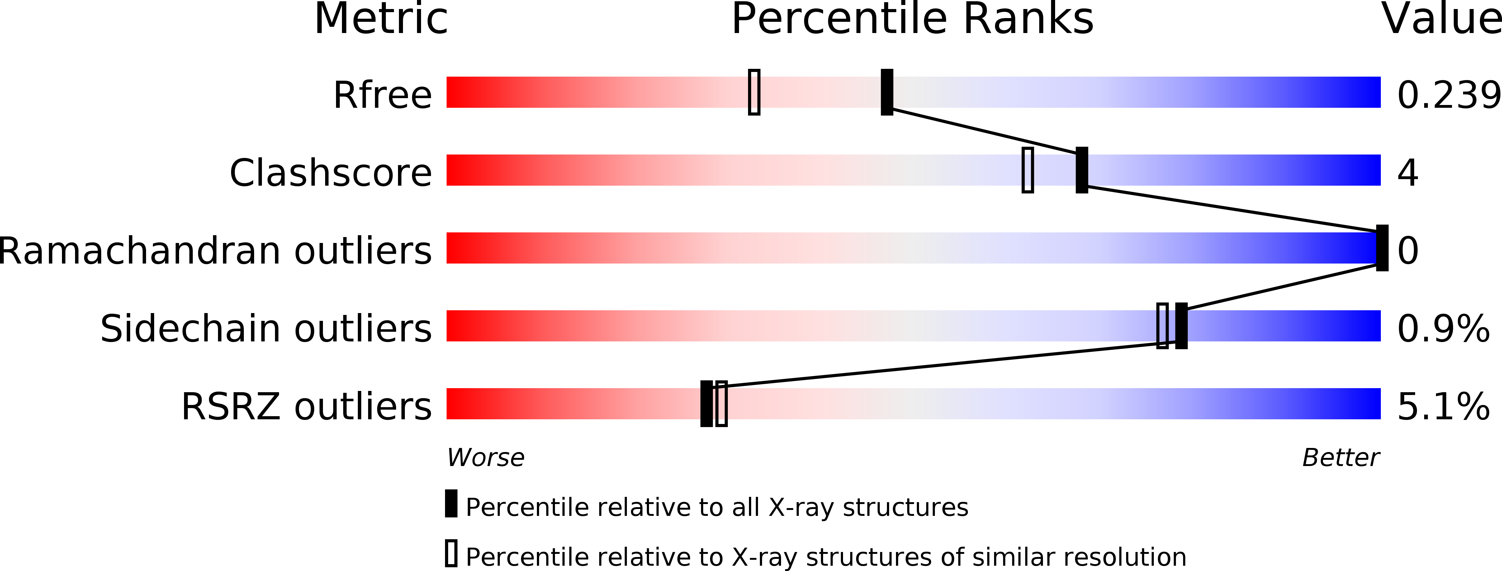
    5OLN

  • PubMed Abstract: 

    Pyrimidine-nucleoside phosphorylase catalyzes the phosphorolytic cleavage of thymidine and uridine with equal activity. Investigation of this protein is essential for anticancer drug design. Here, the structure of this protein from Bacillus subtilis in complex with imidazole and sulfate is reported at 1.9 Å resolution, which is an improvement on the previously reported structure at 2.6 Å resolution. The localization and position of imidazole in the nucleoside-binding site reflects the possible binding of ligands that possess an imidazole ring.


  • Organizational Affiliation

    A. V. Shubnikov Institute of Crystallography, Leninsky Prospect 59, Moscow 119333, Russian Federation.


Macromolecules
Find similar proteins by:  (by identity cutoff)  |  3D Structure
Entity ID: 1
MoleculeChains Sequence LengthOrganismDetailsImage
Pyrimidine-nucleoside phosphorylase
A, B
434Bacillus subtilis subsp. subtilis str. 168Mutation(s): 0 
Gene Names: pdpBSU39400
EC: 2.4.2.2
UniProt
Find proteins for P39142 (Bacillus subtilis (strain 168))
Explore P39142 
Go to UniProtKB:  P39142
Entity Groups  
Sequence Clusters30% Identity50% Identity70% Identity90% Identity95% Identity100% Identity
UniProt GroupP39142
Sequence Annotations
Expand
  • Reference Sequence
Experimental Data & Validation

Experimental Data

  • Method: X-RAY DIFFRACTION
  • Resolution: 1.88 Å
  • R-Value Free: 0.239 
  • R-Value Work: 0.200 
  • R-Value Observed: 0.201 
  • Space Group: P 21 21 21
Unit Cell:
Length ( Å )Angle ( ˚ )
a = 64.368α = 90
b = 93.01β = 90
c = 139.854γ = 90
Software Package:
Software NamePurpose
PHENIXrefinement
SCALAdata scaling
PHASERphasing
PDB_EXTRACTdata extraction
XDSdata reduction

Structure Validation

View Full Validation Report


Entry History & Funding Information

Deposition Data


Funding OrganizationLocationGrant Number
Grant of the President of Russian FederationRussian FederationMK-9246.2016.3.

Revision History  (Full details and data files)

  • Version 1.0: 2018-04-18
    Type: Initial release
  • Version 1.1: 2018-07-11
    Changes: Data collection, Database references, Derived calculations, Structure summary
  • Version 1.2: 2024-01-17
    Changes: Data collection, Database references, Refinement description