5O84

Glutathione S-transferase Tau 23 (partially oxidized)


Experimental Data Snapshot

  • Method: X-RAY DIFFRACTION
  • Resolution: 1.88 Å
  • R-Value Free: 0.216 
  • R-Value Work: 0.181 
  • R-Value Observed: 0.183 

wwPDB Validation   3D Report Full Report


This is version 1.2 of the entry. See complete history


Literature

Disulfide bond formation protects Arabidopsis thaliana glutathione transferase tau 23 from oxidative damage.

Tossounian, M.A.Van Molle, I.Wahni, K.Jacques, S.Gevaert, K.Van Breusegem, F.Vertommen, D.Young, D.Rosado, L.A.Messens, J.

(2018) Biochim Biophys Acta 1862: 775-789

  • DOI: https://doi.org/10.1016/j.bbagen.2017.10.007
  • Primary Citation of Related Structures:  
    5O84, 6EP6, 6EP7

  • PubMed Abstract: 

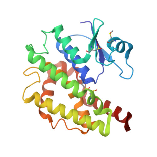
    Glutathione transferases play an important role as detoxifying enzymes. In A. thaliana, elevated levels of reactive oxygen species (ROS), provoked during biotic and abiotic stress, influence the activity of GSTU23. The aim of this study is to determine the impact of oxidative stress on the function and structure of GSTU23. The impact of oxidation on the function of GSTU23 was studied using a glutathione transferase biochemical assay and mass spectrometry. With kinetics, circular dichroism and thermodynamics, we compared reduced with oxidized GSTU23. X-ray crystal structures of GSTU23 visualize the impact of oxidation on methionines and cysteines. In the presence of 100μM H 2 O 2 , oxidation of the methionine side-chain to a sulfoxide is the prominent post-translational modification, which can be reduced by C. diphtheriae MsrA and MsrB. However, increasing the level to 200μM H 2 O 2 results in a reversible intramolecular disulfide between Cys65-Cys110, which is substrate for glutaredoxin. Under these oxidizing conditions, GSTU23 undergoes a structural change and forms a more favourable enzyme-substrate complex to overcome k cat decrease. At lower H 2 O 2 levels (100μM), GSTU23 forms methionine sulfoxides. Specifically, oxidation of Met14, located near the catalytic Ser13, could interfere with both GSH binding and catalytic activation. At higher H 2 O 2 levels (200μM), the Cys65-Cys110 disulfide bond protects other cysteines and also methionines from overoxidation. This study shows the impact of oxidative stress on GSTU23 regulated by methionine sulfoxide reductases and glutaredoxin, and the mechanisms involved in maintaining its catalytic functionality under oxidizing conditions.


  • Organizational Affiliation

    VIB-VUB Center for Structural Biology, B-1050 Brussels, Belgium; Brussels Center for Redox Biology, B-1050 Brussels, Belgium; Structural Biology Brussels, Vrije Universiteit Brussel, B-1050 Brussels, Belgium.


Macromolecules
Find similar proteins by:  (by identity cutoff)  |  3D Structure
Entity ID: 1
MoleculeChains Sequence LengthOrganismDetailsImage
Glutathione S-transferase U23216Arabidopsis thalianaMutation(s): 0 
Gene Names: GSTU23At1g78320F3F9.14
EC: 2.5.1.18
UniProt
Find proteins for Q9M9F1 (Arabidopsis thaliana)
Explore Q9M9F1 
Go to UniProtKB:  Q9M9F1
Entity Groups  
Sequence Clusters30% Identity50% Identity70% Identity90% Identity95% Identity100% Identity
UniProt GroupQ9M9F1
Sequence Annotations
Expand
  • Reference Sequence
Small Molecules
Ligands 5 Unique
IDChains Name / Formula / InChI Key2D Diagram3D Interactions
GOL
Query on GOL

Download Ideal Coordinates CCD File 
F [auth A]GLYCEROL
C3 H8 O3
PEDCQBHIVMGVHV-UHFFFAOYSA-N
EDO
Query on EDO

Download Ideal Coordinates CCD File 
C [auth A],
G [auth A],
H [auth A]
1,2-ETHANEDIOL
C2 H6 O2
LYCAIKOWRPUZTN-UHFFFAOYSA-N
ACT
Query on ACT

Download Ideal Coordinates CCD File 
D [auth A],
I [auth A]
ACETATE ION
C2 H3 O2
QTBSBXVTEAMEQO-UHFFFAOYSA-M
FMT
Query on FMT

Download Ideal Coordinates CCD File 
B [auth A]FORMIC ACID
C H2 O2
BDAGIHXWWSANSR-UHFFFAOYSA-N
MG
Query on MG

Download Ideal Coordinates CCD File 
E [auth A]MAGNESIUM ION
Mg
JLVVSXFLKOJNIY-UHFFFAOYSA-N
Modified Residues  4 Unique
IDChains TypeFormula2D DiagramParent
CSO
Query on CSO
A
L-PEPTIDE LINKINGC3 H7 N O3 SCYS
MHO
Query on MHO
A
L-PEPTIDE LINKINGC5 H11 N O3 SMET
OCS
Query on OCS
A
L-PEPTIDE LINKINGC3 H7 N O5 SCYS
OMT
Query on OMT
A
L-PEPTIDE LINKINGC5 H11 N O4 SMET
Experimental Data & Validation

Experimental Data

  • Method: X-RAY DIFFRACTION
  • Resolution: 1.88 Å
  • R-Value Free: 0.216 
  • R-Value Work: 0.181 
  • R-Value Observed: 0.183 
  • Space Group: C 1 2 1
Unit Cell:
Length ( Å )Angle ( ˚ )
a = 85.165α = 90
b = 50.74β = 112.47
c = 57.167γ = 90
Software Package:
Software NamePurpose
PHENIXrefinement
XDSdata reduction
Aimlessdata scaling
PHASERphasing

Structure Validation

View Full Validation Report


Entry History 

Deposition Data

Revision History  (Full details and data files)

  • Version 1.0: 2017-10-25
    Type: Initial release
  • Version 1.1: 2018-02-14
    Changes: Database references
  • Version 1.2: 2024-01-17
    Changes: Data collection, Database references, Derived calculations, Refinement description