5O0Z

Structure of laspartomycin C in complex with geranyl-phosphate


Experimental Data Snapshot

  • Method: X-RAY DIFFRACTION
  • Resolution: 1.28 Å
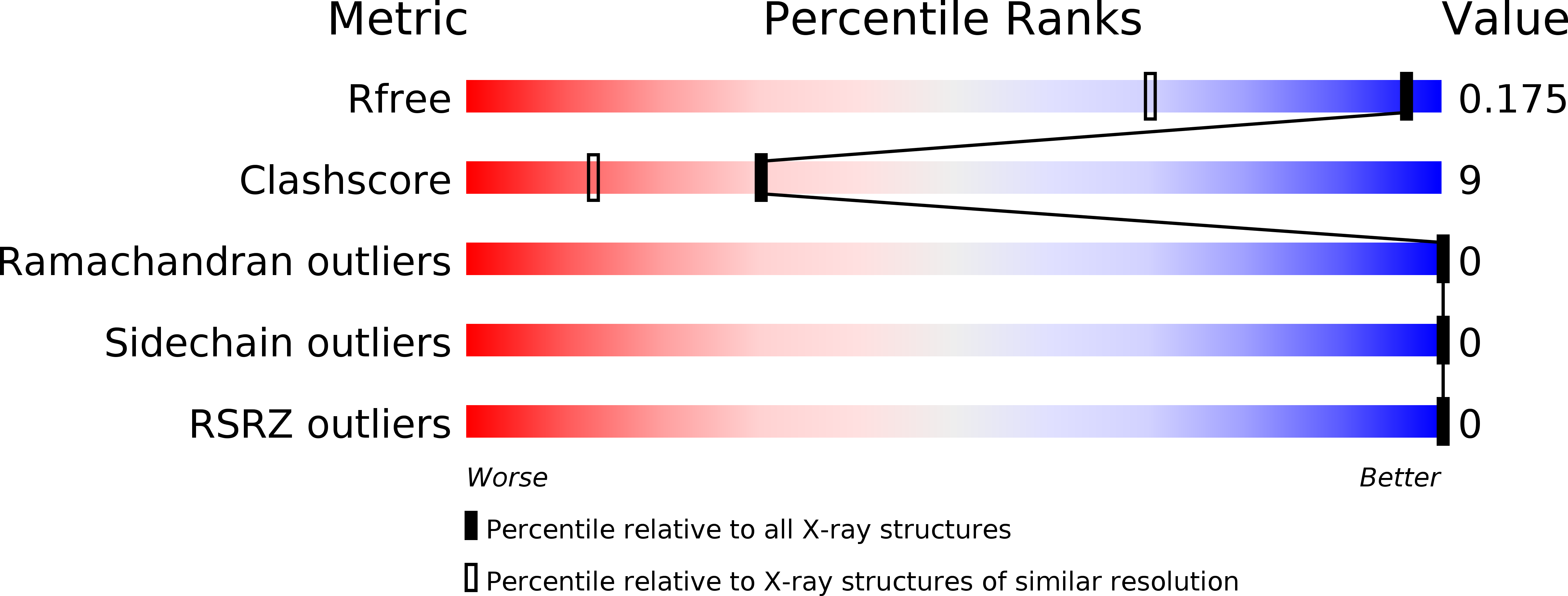
  • R-Value Free: 0.165 
  • R-Value Work: 0.145 
  • R-Value Observed: 0.146 

wwPDB Validation   3D Report Full Report


Ligand Structure Quality Assessment 


This is version 1.3 of the entry. See complete history


Literature

A High-Resolution Crystal Structure that Reveals Molecular Details of Target Recognition by the Calcium-Dependent Lipopeptide Antibiotic Laspartomycin C.

Kleijn, L.H.J.Vlieg, H.C.Wood, T.M.Sastre Torano, J.Janssen, B.J.C.Martin, N.I.

(2017) Angew Chem Int Ed Engl 56: 16546-16549

  • DOI: https://doi.org/10.1002/anie.201709240
  • Primary Citation of Related Structures:  
    5O0Z

  • PubMed Abstract: 

    The calcium-dependent antibiotics (CDAs) are an important emerging class of antibiotics. The crystal structure of the CDA laspartomycin C in complex with calcium and the ligand geranyl-phosphate at a resolution of 1.28 Å is reported. This is the first crystal structure of a CDA bound to its bacterial target. The structure is also the first to be reported for an antibiotic that binds the essential bacterial phospholipid undecaprenyl phosphate (C 55 -P). These structural insights are of great value in the design of antibiotics capable of exploiting this unique bacterial target.


  • Organizational Affiliation

    Department of Chemical Biology & Drug Discovery, Utrecht Institute for Pharmaceutical Sciences, Utrecht University, Universiteitsweg 99, 3584, CG, Utrecht, The Netherlands.


Macromolecules

Find similar proteins by:  Sequence   |   3D Structure  

Entity ID: 1
MoleculeChains Sequence LengthOrganismDetailsImage
Laspartomycin C
A, B
12Streptomyces viridochromogenesMutation(s): 0 
Entity Groups  
Sequence Clusters30% Identity50% Identity70% Identity90% Identity95% Identity100% Identity
Sequence Annotations
Expand
  • Reference Sequence
Small Molecules
Ligands 4 Unique
IDChains Name / Formula / InChI Key2D Diagram3D Interactions
9GB
Query on 9GB

Download Ideal Coordinates CCD File 
C [auth A],
I [auth B]
[(~{E})-3-methylhex-2-enyl] dihydrogen phosphate
C7 H15 O4 P
QFTXOINSFIOLQI-FNORWQNLSA-N
ACY
Query on ACY

Download Ideal Coordinates CCD File 
G [auth A],
H [auth A],
L [auth B]
ACETIC ACID
C2 H4 O2
QTBSBXVTEAMEQO-UHFFFAOYSA-N
CA
Query on CA

Download Ideal Coordinates CCD File 
D [auth A],
E [auth A],
J [auth B],
K [auth B]
CALCIUM ION
Ca
BHPQYMZQTOCNFJ-UHFFFAOYSA-N
CL
Query on CL

Download Ideal Coordinates CCD File 
F [auth A]CHLORIDE ION
Cl
VEXZGXHMUGYJMC-UHFFFAOYSA-M
Modified Residues  1 Unique
IDChains TypeFormula2D DiagramParent
DNP
Query on DNP
A, B
L-PEPTIDE LINKINGC3 H9 N2 O2ALA
Experimental Data & Validation

Experimental Data

  • Method: X-RAY DIFFRACTION
  • Resolution: 1.28 Å
  • R-Value Free: 0.165 
  • R-Value Work: 0.145 
  • R-Value Observed: 0.146 
  • Space Group: P 43 3 2
Unit Cell:
Length ( Å )Angle ( ˚ )
a = 56.933α = 90
b = 56.933β = 90
c = 56.933γ = 90
Software Package:
Software NamePurpose
REFMACrefinement
PDB_EXTRACTdata extraction
iMOSFLMdata reduction
Aimlessdata scaling
SHELXCDphasing

Structure Validation

View Full Validation Report


Ligand Structure Quality Assessment 


Entry History & Funding Information

Deposition Data


Funding OrganizationLocationGrant Number
Netherlands--

Revision History  (Full details and data files)

  • Version 1.0: 2017-11-15
    Type: Initial release
  • Version 1.1: 2017-12-13
    Changes: Database references
  • Version 1.2: 2018-01-03
    Changes: Database references
  • Version 1.3: 2019-10-16
    Changes: Data collection