5NQ7

Crystal structure of laccases from Pycnoporus sanguineus, izoform I


Experimental Data Snapshot

  • Method: X-RAY DIFFRACTION
  • Resolution: 2.75 Å
  • R-Value Free: 0.207 
  • R-Value Work: 0.161 
  • R-Value Observed: 0.164 

wwPDB Validation   3D Report Full Report


This is version 2.1 of the entry. See complete history


Literature

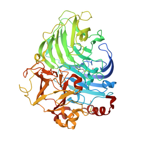
Structural studies of two thermostable laccases from the white-rot fungus Pycnoporus sanguineus.

Orlikowska, M.de J Rostro-Alanis, M.Bujacz, A.Hernandez-Luna, C.Rubio, R.Parra, R.Bujacz, G.

(2018) Int J Biol Macromol 107: 1629-1640

  • DOI: https://doi.org/10.1016/j.ijbiomac.2017.10.024
  • Primary Citation of Related Structures:  
    5NQ7, 5NQ8, 5NQ9

  • PubMed Abstract: 

    Laccases are enzymes that have the ability to catalyze the oxidation of a wide spectrum of phenolic compounds with the four-electron reduction of molecular oxygen to water. The active site of those proteins contains four copper ions, classified into three types. Laccases are interesting enzymes for study from the point of view of their structure, function and application because of their role in lignin degradation. Structural studies of two thermostable laccases produced by the strain Pycnoporus sanguineus CS43 (PsLacI and PsLacII) were performed. Both isoforms of PsLac show high thermal stability, at 60°C and 50°C, respectively, and they remained active at a high concentration of organic solvents. However, PsLacI has a higher thermal and pH stability and tolerance against inhibitors, and is a more efficient catalyst for ABTS and DMP (laccases substrate) than PsLacII. Based on the determined crystal structures we achieved insights into the structural factors relevant for the enzymatic properties of PsLacI and PsLacII. N-glycosylation site Asn354, which is very often present in structures of fungal laccases from other species, was not present in PsLac. This observation may be of particular significance due to the close distance between Asn354 and the substrate-binding pocket. This results in better access to the hydrophobic cavity for a particular substrate. Furthermore, we identified significant differences in the region of substrate-binding pocket, which confer PsLacI a markedly better performance than PsLacII.


  • Organizational Affiliation

    Institute of Technical Biochemistry, Faculty of Biotechnology and Food Sciences, Lodz University of Technology, 90-924 Lodz, Stefanowskiego 4/10, Poland.


Macromolecules
Find similar proteins by:  (by identity cutoff)  |  3D Structure
Entity ID: 1
MoleculeChains Sequence LengthOrganismDetailsImage
Laccase518Trametes sanguineaMutation(s): 0 
EC: 1.10.3.2
UniProt
Find proteins for D7F485 (Trametes sanguinea)
Explore D7F485 
Go to UniProtKB:  D7F485
Entity Groups  
Sequence Clusters30% Identity50% Identity70% Identity90% Identity95% Identity100% Identity
UniProt GroupD7F485
Sequence Annotations
Expand
  • Reference Sequence
Oligosaccharides

Help

Entity ID: 2
MoleculeChains Length2D Diagram Glycosylation3D Interactions
alpha-D-mannopyranose-(1-3)-alpha-D-mannopyranose-(1-6)-[beta-D-mannopyranose-(1-3)]beta-D-mannopyranose-(1-4)-2-acetamido-2-deoxy-beta-D-glucopyranose-(1-4)-2-acetamido-2-deoxy-beta-D-glucopyranose
B
6N-Glycosylation
Glycosylation Resources
GlyTouCan:  G00601VC
GlyCosmos:  G00601VC
GlyGen:  G00601VC
Entity ID: 3
MoleculeChains Length2D Diagram Glycosylation3D Interactions
alpha-D-mannopyranose-(1-3)-[beta-D-mannopyranose-(1-6)]beta-D-mannopyranose-(1-3)-beta-D-mannopyranose-(1-4)-2-acetamido-2-deoxy-beta-D-glucopyranose-(1-4)-2-acetamido-2-deoxy-beta-D-glucopyranose
C
6N-Glycosylation
Glycosylation Resources
GlyTouCan:  G88748KA
GlyCosmos:  G88748KA
GlyGen:  G88748KA
Entity ID: 4
MoleculeChains Length2D Diagram Glycosylation3D Interactions
alpha-D-mannopyranose-(1-3)-beta-D-mannopyranose-(1-4)-2-acetamido-2-deoxy-beta-D-glucopyranose-(1-4)-2-acetamido-2-deoxy-beta-D-glucopyranose
D
4N-Glycosylation
Glycosylation Resources
GlyTouCan:  G81315DD
GlyCosmos:  G81315DD
GlyGen:  G81315DD
Experimental Data & Validation

Experimental Data

  • Method: X-RAY DIFFRACTION
  • Resolution: 2.75 Å
  • R-Value Free: 0.207 
  • R-Value Work: 0.161 
  • R-Value Observed: 0.164 
  • Space Group: P 43 3 2
Unit Cell:
Length ( Å )Angle ( ˚ )
a = 185.55α = 90
b = 185.55β = 90
c = 185.55γ = 90
Software Package:
Software NamePurpose
REFMACrefinement
XDSdata reduction
XDSdata scaling
MOLREPphasing

Structure Validation

View Full Validation Report


Entry History 

Deposition Data

Revision History  (Full details and data files)

  • Version 1.0: 2017-11-01
    Type: Initial release
  • Version 1.1: 2018-01-10
    Changes: Database references
  • Version 2.0: 2020-07-29
    Type: Remediation
    Reason: Carbohydrate remediation
    Changes: Advisory, Atomic model, Data collection, Derived calculations, Structure summary
  • Version 2.1: 2024-01-17
    Changes: Data collection, Database references, Derived calculations, Refinement description, Structure summary