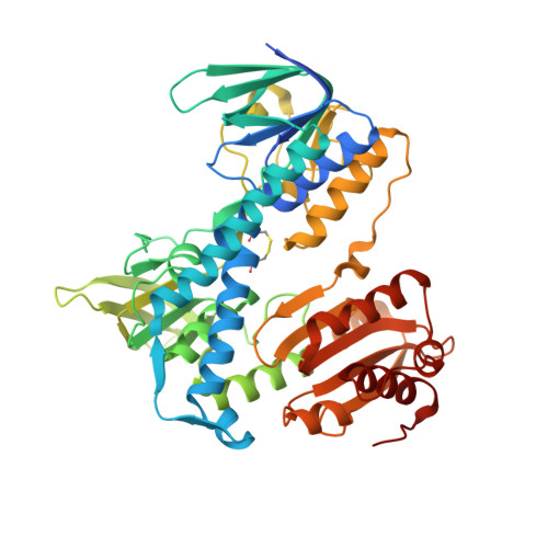
5NHG

Crystal structure of the human dihydrolipoamide dehydrogenase


Experimental Data Snapshot

  • Method: X-RAY DIFFRACTION
  • Resolution: 2.27 Å
  • R-Value Free: 0.210 
  • R-Value Work: 0.180 
  • R-Value Observed: 0.181 

wwPDB Validation   3D Report Full Report


Ligand Structure Quality Assessment 


This is version 1.2 of the entry. See complete history


Literature

Crystal structures of the disease-causing D444V mutant and the relevant wild type human dihydrolipoamide dehydrogenase.

Szabo, E.Mizsei, R.Wilk, P.Zambo, Z.Torocsik, B.Weiss, M.S.Adam-Vizi, V.Ambrus, A.

(2018) Free Radic Biol Med 124: 214-220

  • DOI: https://doi.org/10.1016/j.freeradbiomed.2018.06.008
  • Primary Citation of Related Structures:  
    5J5Z, 5NHG

  • PubMed Abstract: 

    We report the crystal structures of the human (dihydro)lipoamide dehydrogenase (hLADH, hE3) and its disease-causing homodimer interface mutant D444V-hE3 at 2.27 and 1.84 Å resolution, respectively. The wild type structure is a unique uncomplexed, unliganded hE3 structure with the true canonical sequence. Based on the structural information a novel molecular pathomechanism is proposed for the impaired catalytic activity and enhanced capacity for reactive oxygen species generation of the pathogenic mutant. The mechanistic model involves a previously much ignored solvent accessible channel leading to the active site that might be perturbed also by other disease-causing homodimer interface substitutions of this enzyme.


  • Organizational Affiliation

    Department of Medical Biochemistry, MTA-SE Laboratory for Neurobiochemistry, Semmelweis University, H-1094 Budapest, Hungary.


Macromolecules
Find similar proteins by:  (by identity cutoff)  |  3D Structure
Entity ID: 1
MoleculeChains Sequence LengthOrganismDetailsImage
Dihydrolipoyl dehydrogenase, mitochondrial
A, B, C, D, E
A, B, C, D, E, F, G, H
495Homo sapiensMutation(s): 0 
Gene Names: DLDGCSLLADPHE3
EC: 1.8.1.4
UniProt & NIH Common Fund Data Resources
Find proteins for P09622 (Homo sapiens)
Explore P09622 
Go to UniProtKB:  P09622
PHAROS:  P09622
GTEx:  ENSG00000091140 
Entity Groups  
Sequence Clusters30% Identity50% Identity70% Identity90% Identity95% Identity100% Identity
UniProt GroupP09622
Sequence Annotations
Expand
  • Reference Sequence
Small Molecules
Ligands 3 Unique
IDChains Name / Formula / InChI Key2D Diagram3D Interactions
FAD
Query on FAD

Download Ideal Coordinates CCD File 
I [auth A]
L [auth B]
M [auth C]
P [auth D]
Q [auth E]
I [auth A],
L [auth B],
M [auth C],
P [auth D],
Q [auth E],
T [auth F],
V [auth G],
X [auth H]
FLAVIN-ADENINE DINUCLEOTIDE
C27 H33 N9 O15 P2
VWWQXMAJTJZDQX-UYBVJOGSSA-N
BTB
Query on BTB

Download Ideal Coordinates CCD File 
O [auth C]2-[BIS-(2-HYDROXY-ETHYL)-AMINO]-2-HYDROXYMETHYL-PROPANE-1,3-DIOL
C8 H19 N O5
OWMVSZAMULFTJU-UHFFFAOYSA-N
TRS
Query on TRS

Download Ideal Coordinates CCD File 
J [auth A]
K [auth A]
N [auth C]
R [auth E]
S [auth E]
J [auth A],
K [auth A],
N [auth C],
R [auth E],
S [auth E],
U [auth F],
W [auth G]
2-AMINO-2-HYDROXYMETHYL-PROPANE-1,3-DIOL
C4 H12 N O3
LENZDBCJOHFCAS-UHFFFAOYSA-O
Experimental Data & Validation

Experimental Data

  • Method: X-RAY DIFFRACTION
  • Resolution: 2.27 Å
  • R-Value Free: 0.210 
  • R-Value Work: 0.180 
  • R-Value Observed: 0.181 
  • Space Group: P 1
Unit Cell:
Length ( Å )Angle ( ˚ )
a = 60.06α = 83.18
b = 113.54β = 84.75
c = 136.88γ = 81.09
Software Package:
Software NamePurpose
PHENIXrefinement
MxCuBEdata collection
XDSdata scaling
XDSdata reduction
MOLREPphasing

Structure Validation

View Full Validation Report


Ligand Structure Quality Assessment 


Entry History & Funding Information

Deposition Data


Funding OrganizationLocationGrant Number
Hungarian Academy of SciencesHungary02001
Hungarian Scientific Research FundHungary112230
Hungarian Brain Research ProgramHungaryKTIA_13_NAP-A-III/6
Hungarian Academy of SciencesHungaryBolyai Fellowship [to A.A.]
European Molecular Biology OrganizationHungaryShort-term Fellowship [to A.A.]
Helmholtz-Zentrum BerlinGermany16204087-ST, BESSY: 362 [to E.Sz., A.A.]

Revision History  (Full details and data files)

  • Version 1.0: 2018-05-16
    Type: Initial release
  • Version 1.1: 2018-07-04
    Changes: Data collection, Database references
  • Version 1.2: 2024-01-17
    Changes: Data collection, Database references, Refinement description