


Structure of the first cytoplasmic domain of OutF and assembly of the inner-membrane platform proteins of the D. dadantii type II secretion system
Zhang, H., Gu, S.To be published.
Experimental Data Snapshot
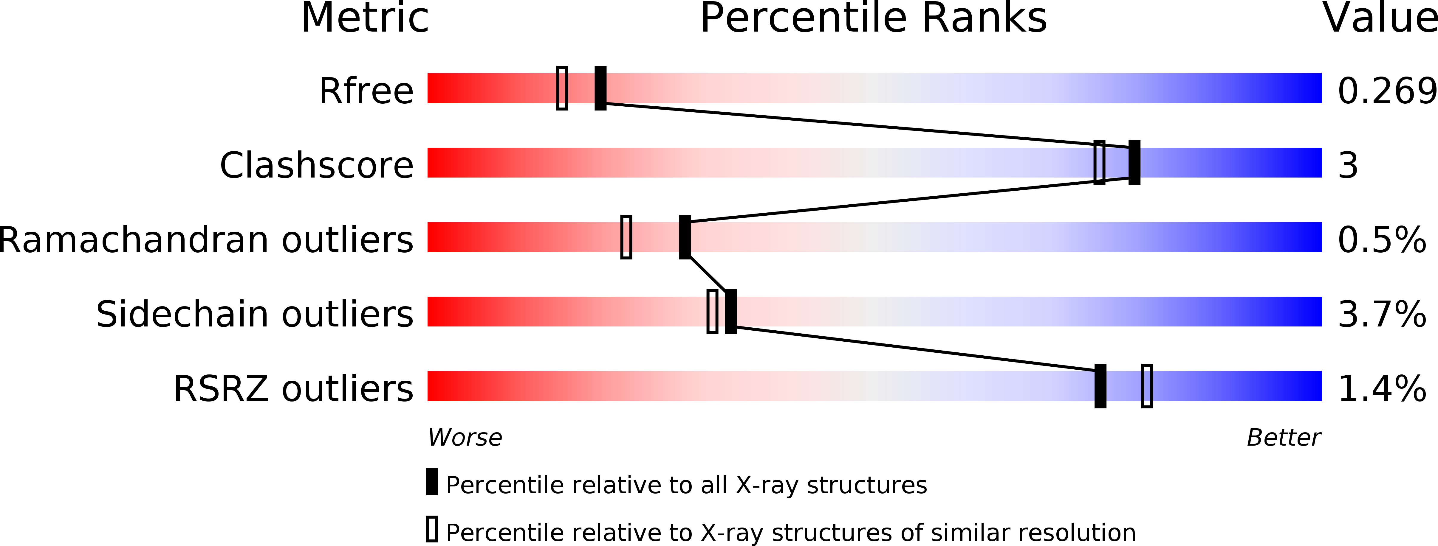
wwPDB Validation 3D Report Full Report
Entity ID: 1 | |||||
|---|---|---|---|---|---|
| Molecule | Chains | Sequence Length | Organism | Details | Image |
| General secretion pathway protein F | 109 | Dickeya dadantii 3937 | Mutation(s): 0 Gene Names: outF, Dda3937_02417 | 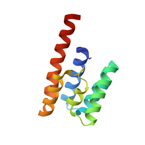 | |
UniProt | |||||
Find proteins for E0SM39 (Dickeya dadantii (strain 3937)) Explore E0SM39 Go to UniProtKB: E0SM39 | |||||
Entity Groups | |||||
| Sequence Clusters | 30% Identity50% Identity70% Identity90% Identity95% Identity100% Identity | ||||
| UniProt Group | E0SM39 | ||||
Sequence AnnotationsExpand | |||||
| |||||
| Length ( Å ) | Angle ( ˚ ) |
|---|---|
| a = 116.175 | α = 90 |
| b = 116.175 | β = 90 |
| c = 47.998 | γ = 90 |
| Software Name | Purpose |
|---|---|
| REFMAC | refinement |
| XDS | data reduction |
| MOLREP | phasing |
| Aimless | data scaling |
RCSB PDB (citation) is hosted by
RCSB PDB is a member of the

