Relaxed and active thin filament structures; a new structural basis for the regulatory mechanism.
Paul, D.M., Squire, J.M., Morris, E.P.(2017) J Struct Biol 197: 365-371
- PubMed: 28161413
- DOI: https://doi.org/10.1016/j.jsb.2017.01.004
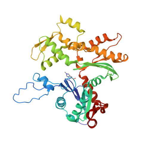
- Primary Citation of Related Structures:
5MVA, 5MVY - PubMed Abstract:
The structures of muscle thin filaments reconstituted using skeletal actin and cardiac troponin and tropomyosin have been determined with and without bound Ca 2+ using electron microscopy and reference-free single particle analysis. The resulting density maps have been fitted with atomic models of actin, tropomyosin and troponin showing that: (i) the polarity of the troponin complex is consistent with our 2009 findings, with large shape changes in troponin between the two states; (ii) without Ca 2+ the tropomyosin pseudo-repeats all lie at almost equivalent positions in the 'blocked' position on actin (over subdomains 1 and 2); (iii) in the active state the tropomyosin pseudo-repeats are all displaced towards subdomains 3 and 4 of actin, but the extent of displacement varies within the regulatory unit depending upon the axial location of the pseudo-repeats with respect to troponin. Individual pseudo-repeats with Ca 2+ bound to troponin can be assigned either to the 'closed' state, a partly activated conformation, or the 'M-state', a fully activated conformation which has previously been thought to occur only when myosin heads bind. These results lead to a modified view of the steric blocking model of thin filament regulation in which cooperative activation is governed by troponin-mediated local interactions of the pseudo-repeats of tropomyosin with actin.
Organizational Affiliation:
Muscle Contraction Group, School of Physiology, Pharmacology and Neuroscience, University of Bristol, Bristol BS8 1TD, UK. Electronic address: Danielle.paul@bristol.ac.uk.