5MTT

Maltodextrin binding protein MalE1 from L. casei BL23 bound to maltotetraose


Experimental Data Snapshot

  • Method: X-RAY DIFFRACTION
  • Resolution: 1.12 Å
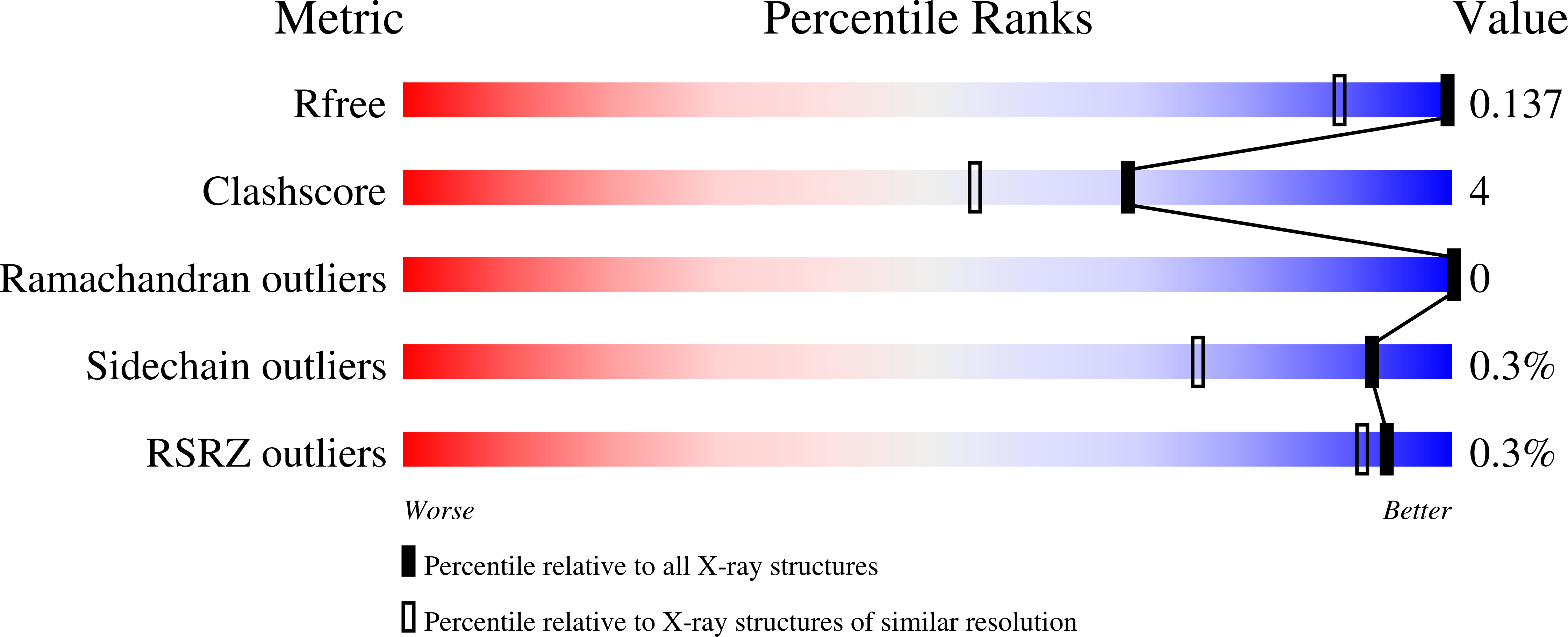
  • R-Value Free: 0.137 
  • R-Value Work: 0.120 
  • R-Value Observed: 0.120 

wwPDB Validation   3D Report Full Report


Ligand Structure Quality Assessment 


This is version 2.0 of the entry. See complete history


Literature

Inducer exclusion in Firmicutes: insights into the regulation of a carbohydrate ATP binding cassette transporter from Lactobacillus casei BL23 by the signal transducing protein P-Ser46-HPr.

Homburg, C.Bommer, M.Wuttge, S.Hobe, C.Beck, S.Dobbek, H.Deutscher, J.Licht, A.Schneider, E.

(2017) Mol Microbiol 105: 25-45

  • DOI: https://doi.org/10.1111/mmi.13680
  • Primary Citation of Related Structures:  
    5M28, 5MK9, 5MKA, 5MKB, 5MTT, 5MTU

  • PubMed Abstract: 

    Catabolite repression is a mechanism that enables bacteria to control carbon utilization. As part of this global regulatory network, components of the phosphoenolpyruvate:carbohydrate phosphotransferase system inhibit the uptake of less favorable sugars when a preferred carbon source such as glucose is available. This process is termed inducer exclusion. In bacteria belonging to the phylum Firmicutes, HPr, phosphorylated at serine 46 (P-Ser46-HPr) is the key player but its mode of action is elusive. To address this question at the level of purified protein components, we have chosen a homolog of the Escherichia coli maltose/maltodextrin ATP-binding cassette transporter from Lactobacillus casei (MalE1-MalF1G1K1 2 ) as a model system. We show that the solute binding protein, MalE1, binds linear and cyclic maltodextrins but not maltose. Crystal structures of MalE1 complexed with these sugars provide a clue why maltose is not a substrate. P-Ser46-HPr inhibited MalE1/maltotetraose-stimulated ATPase activity of the transporter incorporated in proteoliposomes. Furthermore, cross-linking experiments revealed that P-Ser46-HPr contacts the nucleotide-binding subunit, MalK1, in proximity to the Walker A motif. However, P-Ser46-HPr did not block binding of ATP to MalK1. Together, our findings provide first biochemical evidence that P-Ser-HPr arrests the transport cycle by preventing ATP hydrolysis at the MalK1 subunits of the transporter.


  • Organizational Affiliation

    Institut für Biologie/Physiologie der Mikroorganismen, Humboldt-Universität zu Berlin, Berlin, D-10099, Germany.


Macromolecules
Find similar proteins by:  (by identity cutoff)  |  3D Structure
Entity ID: 1
MoleculeChains Sequence LengthOrganismDetailsImage
MalE1379Lacticaseibacillus casei BL23Mutation(s): 0 
Gene Names: malE1
UniProt
Find proteins for B0L7B0 (Lacticaseibacillus casei)
Explore B0L7B0 
Go to UniProtKB:  B0L7B0
Entity Groups  
Sequence Clusters30% Identity50% Identity70% Identity90% Identity95% Identity100% Identity
UniProt GroupB0L7B0
Sequence Annotations
Expand
  • Reference Sequence
Oligosaccharides

Help

Entity ID: 2
MoleculeChains Length2D Diagram Glycosylation3D Interactions
alpha-D-glucopyranose-(1-4)-alpha-D-glucopyranose-(1-4)-alpha-D-glucopyranose-(1-4)-alpha-D-glucopyranose
B
4N/A
Glycosylation Resources
GlyTouCan:  G87171PZ
GlyCosmos:  G87171PZ
GlyGen:  G87171PZ
Small Molecules
Ligands 3 Unique
IDChains Name / Formula / InChI Key2D Diagram3D Interactions
GLC
Query on GLC

Download Ideal Coordinates CCD File 
C [auth A],
D [auth A],
E [auth A],
F [auth A]
alpha-D-glucopyranose
C6 H12 O6
WQZGKKKJIJFFOK-DVKNGEFBSA-N
EDO
Query on EDO

Download Ideal Coordinates CCD File 
G [auth A]
H [auth A]
I [auth A]
J [auth A]
K [auth A]
G [auth A],
H [auth A],
I [auth A],
J [auth A],
K [auth A],
L [auth A],
M [auth A],
N [auth A],
O [auth A],
P [auth A],
Q [auth A]
1,2-ETHANEDIOL
C2 H6 O2
LYCAIKOWRPUZTN-UHFFFAOYSA-N
CL
Query on CL

Download Ideal Coordinates CCD File 
R [auth A],
S [auth A],
T [auth A]
CHLORIDE ION
Cl
VEXZGXHMUGYJMC-UHFFFAOYSA-M
Biologically Interesting Molecules (External Reference) 1 Unique
Experimental Data & Validation

Experimental Data

  • Method: X-RAY DIFFRACTION
  • Resolution: 1.12 Å
  • R-Value Free: 0.137 
  • R-Value Work: 0.120 
  • R-Value Observed: 0.120 
  • Space Group: P 21 21 21
Unit Cell:
Length ( Å )Angle ( ˚ )
a = 53.494α = 90
b = 83.079β = 90
c = 85.036γ = 90
Software Package:
Software NamePurpose
PHENIXrefinement
XDSdata reduction
XDSdata scaling
SHELXDEphasing

Structure Validation

View Full Validation Report


Ligand Structure Quality Assessment 


Entry History & Funding Information

Deposition Data


Funding OrganizationLocationGrant Number
German Research FoundationGermanySFB1078

Revision History  (Full details and data files)

  • Version 1.0: 2017-07-05
    Type: Initial release
  • Version 2.0: 2020-07-29
    Type: Remediation
    Reason: Carbohydrate remediation
    Changes: Advisory, Atomic model, Author supporting evidence, Data collection, Derived calculations, Structure summary