5MOL

Human IgE-Fc crystal structure


Experimental Data Snapshot

  • Method: X-RAY DIFFRACTION
  • Resolution: 1.75 Å
  • R-Value Free: 0.232 
  • R-Value Work: 0.197 
  • R-Value Observed: 0.199 

wwPDB Validation   3D Report Full Report


This is version 2.1 of the entry. See complete history


Literature

Thermal sensitivity and flexibility of the C epsilon 3 domains in immunoglobulin E.

Dore, K.A.Davies, A.M.Drinkwater, N.Beavil, A.J.McDonnell, J.M.Sutton, B.J.

(2017) Biochim Biophys Acta 1865: 1336-1347

  • DOI: https://doi.org/10.1016/j.bbapap.2017.08.005
  • Primary Citation of Related Structures:  
    5MOI, 5MOJ, 5MOK, 5MOL

  • PubMed Abstract: 

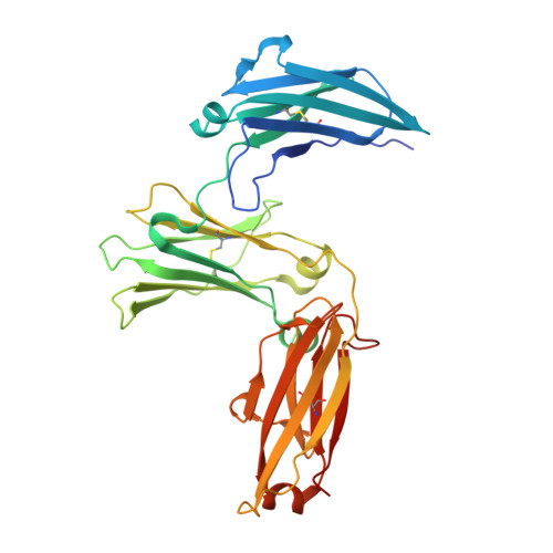
    Immunoglobulin E (IgE) is the antibody that plays a central role in the mechanisms of allergic diseases such as asthma. Interactions with its receptors, FcεRI on mast cells and CD23 on B cells, are mediated by the Fc region, a dimer of the Cε2, Cε3 and Cε4 domains. A sub-fragment lacking the Cε2 domains, Fcε3-4, also binds to both receptors, although receptor binding almost exclusively involves the Cε3 domains. This domain also contains the N-linked glycosylation site conserved in other isotypes. We report here the crystal structures of IgE-Fc and Fcε3-4 at the highest resolutions yet determined, 1.75Å and 2.0Å respectively, revealing unprecedented detail regarding the carbohydrate and its interactions with protein domains. Analysis of the crystallographic B-factors of these, together with all earlier IgE-Fc and Fcε3-4 structures, shows that the Cε3 domains exhibit the greatest intrinsic flexibility and quaternary structural variation within IgE-Fc. Intriguingly, both well-ordered carbohydrate and disordered polypeptide can be seen within the same Cε3 domain. A simplified method for comparing the quaternary structures of the Cε3 domains in free and receptor-bound IgE-Fc structures is presented, which clearly delineates the FcεRI and CD23 bound states. Importantly, differential scanning fluorimetric analysis of IgE-Fc and Fcε3-4 identifies Cε3 as the domain most susceptible to thermally-induced unfolding, and responsible for the characteristically low melting temperature of IgE.


  • Organizational Affiliation

    King's College London, Randall Division of Cell and Molecular Biophysics, New Hunt's House, London SE1 1UL, United Kingdom; Medical Research Council & Asthma UK Centre in Allergic Mechanisms of Asthma, London, United Kingdom.


Macromolecules
Find similar proteins by:  (by identity cutoff)  |  3D Structure
Entity ID: 1
MoleculeChains Sequence LengthOrganismDetailsImage
Ig epsilon chain C region
A, B
327Homo sapiensMutation(s): 0 
Gene Names: IGHE
UniProt & NIH Common Fund Data Resources
Find proteins for P01854 (Homo sapiens)
Explore P01854 
Go to UniProtKB:  P01854
PHAROS:  P01854
GTEx:  ENSG00000211891 
Entity Groups  
Sequence Clusters30% Identity50% Identity70% Identity90% Identity95% Identity100% Identity
UniProt GroupP01854
Sequence Annotations
Expand
  • Reference Sequence
Oligosaccharides

Help

Entity ID: 2
MoleculeChains Length2D Diagram Glycosylation3D Interactions
alpha-D-mannopyranose-(1-6)-alpha-D-mannopyranose-(1-6)-beta-D-mannopyranose-(1-4)-2-acetamido-2-deoxy-beta-D-glucopyranose-(1-4)-2-acetamido-2-deoxy-beta-D-glucopyranose
C
5N-Glycosylation
Glycosylation Resources
GlyTouCan:  G94626GC
GlyCosmos:  G94626GC
GlyGen:  G94626GC
Entity ID: 3
MoleculeChains Length2D Diagram Glycosylation3D Interactions
alpha-D-mannopyranose-(1-3)-[alpha-D-mannopyranose-(1-6)]alpha-D-mannopyranose-(1-6)-[alpha-D-mannopyranose-(1-3)]beta-D-mannopyranose-(1-4)-2-acetamido-2-deoxy-beta-D-glucopyranose-(1-4)-2-acetamido-2-deoxy-beta-D-glucopyranose
D
7N-Glycosylation
Glycosylation Resources
GlyTouCan:  G55220VL
GlyCosmos:  G55220VL
GlyGen:  G55220VL
Small Molecules
Ligands 4 Unique
IDChains Name / Formula / InChI Key2D Diagram3D Interactions
AE3
Query on AE3

Download Ideal Coordinates CCD File 
K [auth B]2-(2-ETHOXYETHOXY)ETHANOL
C6 H14 O3
XXJWXESWEXIICW-UHFFFAOYSA-N
PG0
Query on PG0

Download Ideal Coordinates CCD File 
E [auth A],
J [auth B]
2-(2-METHOXYETHOXY)ETHANOL
C5 H12 O3
SBASXUCJHJRPEV-UHFFFAOYSA-N
PEG
Query on PEG

Download Ideal Coordinates CCD File 
I [auth A],
N [auth B]
DI(HYDROXYETHYL)ETHER
C4 H10 O3
MTHSVFCYNBDYFN-UHFFFAOYSA-N
EDO
Query on EDO

Download Ideal Coordinates CCD File 
F [auth A],
G [auth A],
H [auth A],
L [auth B],
M [auth B]
1,2-ETHANEDIOL
C2 H6 O2
LYCAIKOWRPUZTN-UHFFFAOYSA-N
Experimental Data & Validation

Experimental Data

  • Method: X-RAY DIFFRACTION
  • Resolution: 1.75 Å
  • R-Value Free: 0.232 
  • R-Value Work: 0.197 
  • R-Value Observed: 0.199 
  • Space Group: P 21 21 2
Unit Cell:
Length ( Å )Angle ( ˚ )
a = 130.674α = 90
b = 75.782β = 90
c = 79.817γ = 90
Software Package:
Software NamePurpose
PHENIXrefinement
DIALSdata reduction
Aimlessdata scaling
PHASERphasing

Structure Validation

View Full Validation Report


Entry History & Funding Information

Deposition Data


Funding OrganizationLocationGrant Number
Medical Research Council (United Kingdom)United KingdomG1100090

Revision History  (Full details and data files)

  • Version 1.0: 2018-01-10
    Type: Initial release
  • Version 2.0: 2020-07-29
    Type: Remediation
    Reason: Carbohydrate remediation
    Changes: Atomic model, Data collection, Derived calculations, Structure summary
  • Version 2.1: 2024-01-17
    Changes: Data collection, Database references, Derived calculations, Refinement description, Structure summary