


Productive ternary complexes of D-2-hydroxyacid dehydrogenase provide insights into the chiral specificity of its reaction mechanism
Domenech Perez, J., Pramanpol, N., Baker, P.J., Bisson, C., Harding, S.E., Rice, D.W., Ferrer, J.To be published.
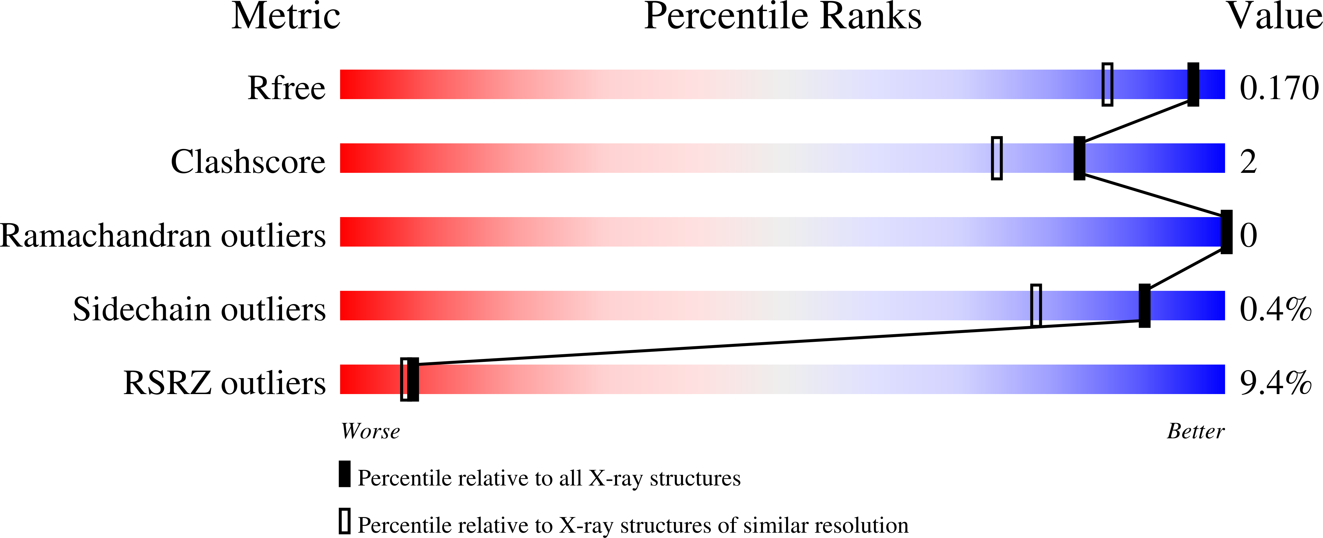
Experimental Data Snapshot
Entity ID: 1 | |||||
|---|---|---|---|---|---|
| Molecule | Chains | Sequence Length | Organism | Details | Image |
| D-2-hydroxyacid dehydrogenase | 308 | Haloferax mediterranei ATCC 33500 | Mutation(s): 0 Gene Names: ddh, serA5, HFX_2024 EC: 1.1.1 | 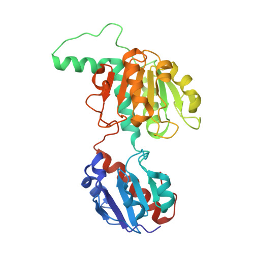 | |
UniProt | |||||
Find proteins for Q2VEQ7 (Haloferax mediterranei (strain ATCC 33500 / DSM 1411 / JCM 8866 / NBRC 14739 / NCIMB 2177 / R-4)) Explore Q2VEQ7 Go to UniProtKB: Q2VEQ7 | |||||
Entity Groups | |||||
| Sequence Clusters | 30% Identity50% Identity70% Identity90% Identity95% Identity100% Identity | ||||
| UniProt Group | Q2VEQ7 | ||||
Sequence AnnotationsExpand | |||||
| |||||
| Ligands 4 Unique | |||||
|---|---|---|---|---|---|
| ID | Chains | Name / Formula / InChI Key | 2D Diagram | 3D Interactions | |
| NAP Query on NAP | BA [auth B], P [auth A] | NADP NICOTINAMIDE-ADENINE-DINUCLEOTIDE PHOSPHATE C21 H28 N7 O17 P3 XJLXINKUBYWONI-NNYOXOHSSA-N |  | ||
| 7N5 Query on 7N5 | AA [auth B], O [auth A] | 2-Ketohexanoic acid C6 H10 O3 XNIHZNNZJHYHLC-UHFFFAOYSA-N |  | ||
| SO4 Query on SO4 | CA [auth B], Q [auth A] | SULFATE ION O4 S QAOWNCQODCNURD-UHFFFAOYSA-L |  | ||
| MG Query on MG | C [auth A] D [auth A] E [auth A] F [auth A] G [auth A] | MAGNESIUM ION Mg JLVVSXFLKOJNIY-UHFFFAOYSA-N |  | ||
| Length ( Å ) | Angle ( ˚ ) |
|---|---|
| a = 62.53 | α = 90 |
| b = 87.4 | β = 96.34 |
| c = 66.1 | γ = 90 |
| Software Name | Purpose |
|---|---|
| REFMAC | refinement |
| iMOSFLM | data reduction |
| Aimless | data scaling |
| Coot | model building |
| Funding Organization | Location | Grant Number |
|---|---|---|
| University of Alicante | Spain | VIGROB046 |
| Biotechnology and Biological Sciences Research Council | United Kingdom | -- |
| Wellcome Trust | United Kingdom | -- |
| Wolfson Foundation | United Kingdom | -- |
RCSB PDB (citation) is hosted by
RCSB PDB is a member of the

