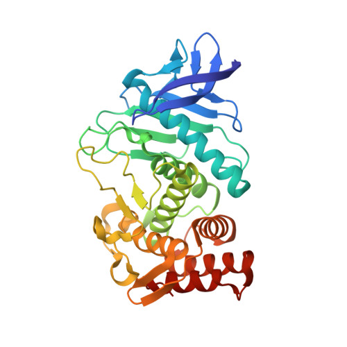
5MA7

Structure of thermolysin in complex with inhibitor (JC306).


Experimental Data Snapshot

  • Method: X-RAY DIFFRACTION
  • Resolution: 1.30 Å
  • R-Value Free: 0.139 
  • R-Value Work: 0.112 
  • R-Value Observed: 0.113 

wwPDB Validation   3D Report Full Report


Ligand Structure Quality Assessment 


This is version 1.1 of the entry. See complete history


Literature

How Nothing Boosts Affinity: Hydrophobic Ligand Binding to the Virtually Vacated S1' Pocket of Thermolysin.

Krimmer, S.G.Cramer, J.Schiebel, J.Heine, A.Klebe, G.

(2017) J Am Chem Soc 139: 10419-10431

  • DOI: https://doi.org/10.1021/jacs.7b05028
  • Primary Citation of Related Structures:  
    5LVD, 5M5F, 5M69, 5M9W, 5MA7

  • PubMed Abstract: 

    We investigated the hydration state of the deep, well-accessible hydrophobic S 1 ' specificity pocket of the metalloprotease thermolysin with purposefully designed ligands using high-resolution crystallography and isothermal titration calorimetry. The S 1 ' pocket is known to recognize selectively a very stringent set of aliphatic side chains such as valine, leucine, and isoleucine of putative substrates. We engineered a weak-binding ligand covering the active site of the protease without addressing the S 1 ' pocket, thus transforming it into an enclosed cavity. Its sustained accessibility could be proved by accommodating noble gas atoms into the pocket in the crystalline state. The topology and electron content of the enclosed pocket with a volume of 141 Å 3 were analyzed using an experimental MAD-phased electron density map that was calibrated to an absolute electron number scale, enabling access to the total electron content within the cavity. Our analysis indicates that the S 1 ' pocket is virtually vacated, thus free of any water molecules. The thermodynamic signature of the reduction of the void within the pocket by growing aliphatic P 1 ' substituents (H, Me, iPr, iBu) reveals a dramatic, enthalpy-dominated gain in free energy of binding resulting in a factor of 41 000 in K d for the H-to-iBu transformation. Substituents placing polar decoy groups into the pocket to capture putatively present water molecules could not collect any evidence for a bound solvent molecule.


  • Organizational Affiliation

    Department of Pharmaceutical Chemistry, University of Marburg , Marbacher Weg 6, 35032 Marburg, Germany.


Macromolecules
Find similar proteins by:  (by identity cutoff)  |  3D Structure
Entity ID: 1
MoleculeChains Sequence LengthOrganismDetailsImage
ThermolysinA [auth E]316Bacillus thermoproteolyticusMutation(s): 0 
EC: 3.4.24.27
UniProt
Find proteins for P00800 (Bacillus thermoproteolyticus)
Explore P00800 
Go to UniProtKB:  P00800
Entity Groups  
Sequence Clusters30% Identity50% Identity70% Identity90% Identity95% Identity100% Identity
UniProt GroupP00800
Sequence Annotations
Expand
  • Reference Sequence
Small Molecules
Ligands 5 Unique
IDChains Name / Formula / InChI Key2D Diagram3D Interactions
7K0
Query on 7K0

Download Ideal Coordinates CCD File 
M [auth E](2~{S})-2-[[(2~{S})-3-azanyl-2-[[oxidanyl(phenylmethoxycarbonylaminomethyl)phosphoryl]amino]propanoyl]amino]-4-methyl-pentanoic acid
C18 H29 N4 O7 P
XZHIMTMEWJSRRL-GJZGRUSLSA-N
GOL
Query on GOL

Download Ideal Coordinates CCD File 
G [auth E],
H [auth E]
GLYCEROL
C3 H8 O3
PEDCQBHIVMGVHV-UHFFFAOYSA-N
DMS
Query on DMS

Download Ideal Coordinates CCD File 
I [auth E],
J [auth E],
K [auth E],
L [auth E]
DIMETHYL SULFOXIDE
C2 H6 O S
IAZDPXIOMUYVGZ-UHFFFAOYSA-N
ZN
Query on ZN

Download Ideal Coordinates CCD File 
B [auth E]ZINC ION
Zn
PTFCDOFLOPIGGS-UHFFFAOYSA-N
CA
Query on CA

Download Ideal Coordinates CCD File 
C [auth E],
D [auth E],
E,
F [auth E]
CALCIUM ION
Ca
BHPQYMZQTOCNFJ-UHFFFAOYSA-N
Binding Affinity Annotations 
IDSourceBinding Affinity
7K0 Binding MOAD:  5MA7 Kd: 3.36e+5 (nM) from 1 assay(s)
Experimental Data & Validation

Experimental Data

  • Method: X-RAY DIFFRACTION
  • Resolution: 1.30 Å
  • R-Value Free: 0.139 
  • R-Value Work: 0.112 
  • R-Value Observed: 0.113 
  • Space Group: P 61 2 2
Unit Cell:
Length ( Å )Angle ( ˚ )
a = 92.754α = 90
b = 92.754β = 90
c = 129.766γ = 120
Software Package:
Software NamePurpose
PHENIXrefinement
XDSdata reduction
XDSdata scaling
PHASERphasing

Structure Validation

View Full Validation Report


Ligand Structure Quality Assessment 


Entry History & Funding Information

Deposition Data


Funding OrganizationLocationGrant Number
European Research CouncilGermany268145-DrugProfilBind

Revision History  (Full details and data files)

  • Version 1.0: 2017-08-16
    Type: Initial release
  • Version 1.1: 2024-01-17
    Changes: Data collection, Database references, Derived calculations, Refinement description