5LWD

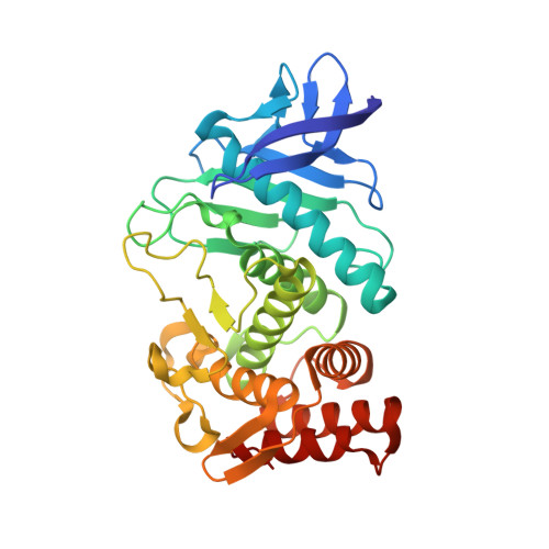
Thermolysin in complex with inhibitor (JC96)


Experimental Data Snapshot

  • Method: X-RAY DIFFRACTION
  • Resolution: 1.23 Å
  • R-Value Free: 0.123 
  • R-Value Work: 0.104 
  • R-Value Observed: 0.105 

wwPDB Validation   3D Report Full Report


This is version 1.2 of the entry. See complete history


Literature

Elucidating the Origin of Long Residence Time Binding for Inhibitors of the Metalloprotease Thermolysin.

Cramer, J.Krimmer, S.G.Fridh, V.Wulsdorf, T.Karlsson, R.Heine, A.Klebe, G.

(2017) ACS Chem Biol 12: 225-233

  • DOI: https://doi.org/10.1021/acschembio.6b00979
  • Primary Citation of Related Structures:  
    5LIF, 5LWD

  • PubMed Abstract: 

    Kinetic parameters of protein-ligand interactions are progressively acknowledged as valuable information for rational drug discovery. However, a targeted optimization of binding kinetics is not easy to achieve, and further systematic studies are necessary to increase the understanding about molecular mechanisms involved. We determined association and dissociation rate constants for 17 inhibitors of the metalloprotease thermolysin by surface plasmon resonance spectroscopy and correlated kinetic data with high-resolution crystal structures in complex with the protein. From the structure-kinetics relationship, we conclude that the strength of interaction with Asn112 correlates with the rate-limiting step of dissociation. This residue is located at the beginning of a β-strand motif that lines the binding cleft and is commonly believed to align a substrate for catalysis. A reduced mobility of the Asn112 side chain owing to an enhanced engagement in charge-assisted hydrogen bonds prevents the conformational adjustment associated with ligand release and transformation of the enzyme to its open state. This hypothesis is supported by kinetic data of ZF P LA, a known pseudopeptidic inhibitor of thermolysin, which blocks the conformational transition of Asn112. Interference with this retrograde induced-fit mechanism results in variation of the residence time of thermolysin inhibitors by a factor of 74 000. The high conservation of this structural motif within the M4 and M13 metalloprotease families underpins the importance of this feature and has significant implications for drug discovery.


  • Organizational Affiliation

    Institute of Pharmaceutical Chemistry, University of Marburg , Marbacher Weg 6, 35032 Marburg, Germany.


Macromolecules
Find similar proteins by:  (by identity cutoff)  |  3D Structure
Entity ID: 1
MoleculeChains Sequence LengthOrganismDetailsImage
ThermolysinA [auth E]316Bacillus thermoproteolyticusMutation(s): 0 
EC: 3.4.24.27
UniProt
Find proteins for P00800 (Bacillus thermoproteolyticus)
Explore P00800 
Go to UniProtKB:  P00800
Entity Groups  
Sequence Clusters30% Identity50% Identity70% Identity90% Identity95% Identity100% Identity
UniProt GroupP00800
Sequence Annotations
Expand
  • Reference Sequence
Small Molecules
Ligands 5 Unique
IDChains Name / Formula / InChI Key2D Diagram3D Interactions
79E
Query on 79E

Download Ideal Coordinates CCD File 
L [auth E]JC96
C21 H35 N4 O6 P
DYUJUGYKDAQNDY-ROUUACIJSA-N
GOL
Query on GOL

Download Ideal Coordinates CCD File 
J [auth E],
K [auth E]
GLYCEROL
C3 H8 O3
PEDCQBHIVMGVHV-UHFFFAOYSA-N
DMS
Query on DMS

Download Ideal Coordinates CCD File 
G [auth E],
H [auth E],
I [auth E]
DIMETHYL SULFOXIDE
C2 H6 O S
IAZDPXIOMUYVGZ-UHFFFAOYSA-N
ZN
Query on ZN

Download Ideal Coordinates CCD File 
B [auth E]ZINC ION
Zn
PTFCDOFLOPIGGS-UHFFFAOYSA-N
CA
Query on CA

Download Ideal Coordinates CCD File 
C [auth E],
D [auth E],
E,
F [auth E]
CALCIUM ION
Ca
BHPQYMZQTOCNFJ-UHFFFAOYSA-N
Binding Affinity Annotations 
IDSourceBinding Affinity
79E Binding MOAD:  5LWD Kd: 570 (nM) from 1 assay(s)
Experimental Data & Validation

Experimental Data

  • Method: X-RAY DIFFRACTION
  • Resolution: 1.23 Å
  • R-Value Free: 0.123 
  • R-Value Work: 0.104 
  • R-Value Observed: 0.105 
  • Space Group: P 61 2 2
Unit Cell:
Length ( Å )Angle ( ˚ )
a = 92.795α = 90
b = 92.795β = 90
c = 130.629γ = 120
Software Package:
Software NamePurpose
PHENIXrefinement
XDSdata reduction
XDSdata scaling
PHASERphasing

Structure Validation

View Full Validation Report


Entry History & Funding Information

Deposition Data


Funding OrganizationLocationGrant Number
European Research Council (ERC)Germany268145-DrugProfilBind

Revision History  (Full details and data files)

  • Version 1.0: 2016-12-21
    Type: Initial release
  • Version 1.1: 2017-02-01
    Changes: Database references
  • Version 1.2: 2024-01-17
    Changes: Data collection, Database references, Derived calculations, Refinement description