Structure of the Branched-chain Amino Acid and GTP-sensing Global Regulator, CodY, from Bacillus subtilis.
Levdikov, V.M., Blagova, E., Young, V.L., Belitsky, B.R., Lebedev, A., Sonenshein, A.L., Wilkinson, A.J.(2017) J Biol Chem 292: 2714-2728
- PubMed: 28011634
- DOI: https://doi.org/10.1074/jbc.M116.754309
- Primary Citation of Related Structures:
5LNH, 5LOE, 5LOJ, 5LOO - PubMed Abstract:
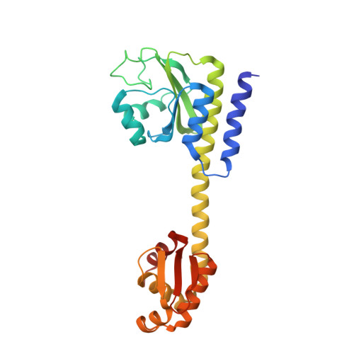
CodY is a branched-chain amino acid (BCAA) and GTP sensor and a global regulator of transcription in low G + C Gram-positive bacteria. It controls the expression of over 100 genes and operons, principally by repressing during growth genes whose products are required for adaptations to nutrient limitation. However, the mechanism by which BCAA binding regulates transcriptional changes is not clear. It is known that CodY consists of a GAF (c G MP-stimulated phosphodiesterases, a denylate cyclases, F hlA) domain that binds BCAAs and a winged helix-turn-helix (wHTH) domain that binds to DNA, but the way in which these domains interact and the structural basis of the BCAA dependence of this interaction are unknown. To gain new insights, we determined the crystal structure of unliganded CodY from Bacillus subtilis revealing a 10-turn α-helix linking otherwise discrete GAF and wHTH domains. The structure of CodY in complex with isoleucine revealed a reorganized GAF domain. In both complexes CodY was tetrameric. Size exclusion chromatography with multiangle laser light scattering (SEC-MALLS) experiments showed that CodY is a dimer at concentrations found in bacterial cells. Comparison of structures of dimers of unliganded CodY and CodY-Ile derived from the tetramers showed a splaying of the wHTH domains when Ile was bound; splaying is likely to account for the increased affinity of Ile-bound CodY for DNA. Electrophoretic mobility shift and SEC-MALLS analyses of CodY binding to 19-36-bp operator fragments are consistent with isoleucine-dependent binding of two CodY dimers per duplex. The implications of these observations for effector control of CodY activity are discussed.
Organizational Affiliation:
From the Structural Biology Laboratory, Department of Chemistry, University of York, York YO10 5DD, United Kingdom.