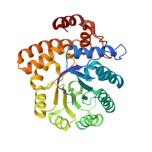
5KZE

N-acetylneuraminate lyase from methicillin-resistant Staphylococcus aureus


Experimental Data Snapshot

  • Method: X-RAY DIFFRACTION
  • Resolution: 1.74 Å
  • R-Value Free: 0.203 
  • R-Value Work: 0.141 
  • R-Value Observed: 0.144 

wwPDB Validation   3D Report Full Report


This is version 1.1 of the entry. See complete history


Literature

Structure and inhibition of N-acetylneuraminate lyase from methicillin-resistant Staphylococcus aureus.

North, R.A.Watson, A.J.Pearce, F.G.Muscroft-Taylor, A.C.Friemann, R.Fairbanks, A.J.Dobson, R.C.

(2016) FEBS Lett 590: 4414-4428

  • DOI: https://doi.org/10.1002/1873-3468.12462
  • Primary Citation of Related Structures:  
    5KZD, 5KZE

  • PubMed Abstract: 

    N-Acetylneuraminate lyase is the first committed enzyme in the degradation of sialic acid by bacterial pathogens. In this study, we analyzed the kinetic parameters of N-acetylneuraminate lyase from methicillin-resistant Staphylococcus aureus (MRSA). We determined that the enzyme has a relatively high K M of 3.2 mm, suggesting that flux through the catabolic pathway is likely to be controlled by this enzyme. Our data indicate that sialic acid alditol, a known inhibitor of N-acetylneuraminate lyase enzymes, is a stronger inhibitor of MRSA N-acetylneuraminate lyase than of Clostridium perfringens N-acetylneuraminate lyase. Our analysis of the crystal structure of ligand-free and 2R-sialic acid alditol-bound MRSA N-acetylneuraminate lyase suggests that subtle dynamic differences in solution and/or altered binding interactions within the active site may account for species-specific inhibition.


  • Organizational Affiliation

    Biomolecular Interaction Centre, School of Biological Sciences, University of Canterbury, Christchurch, New Zealand.


Macromolecules
Find similar proteins by:  (by identity cutoff)  |  3D Structure
Entity ID: 1
MoleculeChains Sequence LengthOrganismDetailsImage
N-acetylneuraminate lyase
A, B, C, D, E
A, B, C, D, E, F, G, H
293Staphylococcus aureus subsp. aureus USA300Mutation(s): 0 
Gene Names: nanASAUSA300_0315
EC: 4.1.3.3
UniProt
Find proteins for Q2G160 (Staphylococcus aureus (strain NCTC 8325 / PS 47))
Explore Q2G160 
Go to UniProtKB:  Q2G160
Entity Groups  
Sequence Clusters30% Identity50% Identity70% Identity90% Identity95% Identity100% Identity
UniProt GroupQ2G160
Sequence Annotations
Expand
  • Reference Sequence
Small Molecules
Ligands 2 Unique
IDChains Name / Formula / InChI Key2D Diagram3D Interactions
SO4
Query on SO4

Download Ideal Coordinates CCD File 
I [auth A]
K [auth B]
M [auth C]
O [auth D]
Q [auth E]
I [auth A],
K [auth B],
M [auth C],
O [auth D],
Q [auth E],
S [auth F],
U [auth G],
W [auth H]
SULFATE ION
O4 S
QAOWNCQODCNURD-UHFFFAOYSA-L
GOL
Query on GOL

Download Ideal Coordinates CCD File 
J [auth A]
L [auth B]
N [auth C]
P [auth D]
R [auth E]
J [auth A],
L [auth B],
N [auth C],
P [auth D],
R [auth E],
T [auth F],
V [auth G],
X [auth H]
GLYCEROL
C3 H8 O3
PEDCQBHIVMGVHV-UHFFFAOYSA-N
Experimental Data & Validation

Experimental Data

  • Method: X-RAY DIFFRACTION
  • Resolution: 1.74 Å
  • R-Value Free: 0.203 
  • R-Value Work: 0.141 
  • R-Value Observed: 0.144 
  • Space Group: P 1 21 1
Unit Cell:
Length ( Å )Angle ( ˚ )
a = 80.11α = 90
b = 130.54β = 90.01
c = 108.52γ = 90
Software Package:
Software NamePurpose
PHENIXrefinement
SCALAdata scaling
PDB_EXTRACTdata extraction
iMOSFLMdata reduction
PHASERphasing

Structure Validation

View Full Validation Report


Entry History 

Deposition Data

Revision History  (Full details and data files)

  • Version 1.0: 2017-01-11
    Type: Initial release
  • Version 1.1: 2023-10-04
    Changes: Data collection, Database references, Refinement description