5KOM

The crystal structure of fluoride channel Fluc Ec2 F83I Mutant


Experimental Data Snapshot

  • Method: X-RAY DIFFRACTION
  • Resolution: 2.69 Å
  • R-Value Free: 0.230 
  • R-Value Work: 0.217 
  • R-Value Observed: 0.218 

wwPDB Validation   3D Report Full Report


Ligand Structure Quality Assessment 


This is version 1.4 of the entry. See complete history


Literature

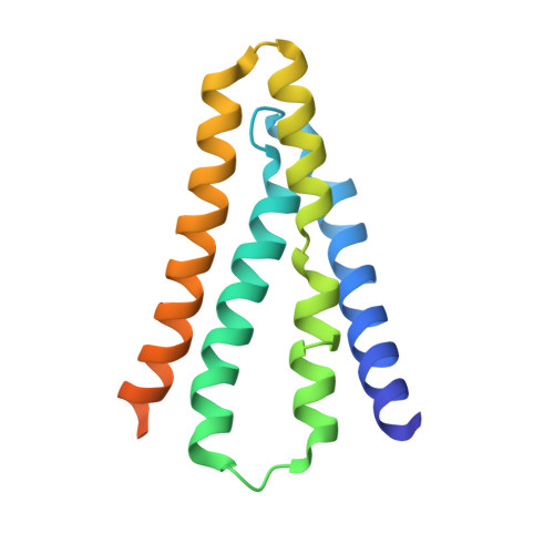
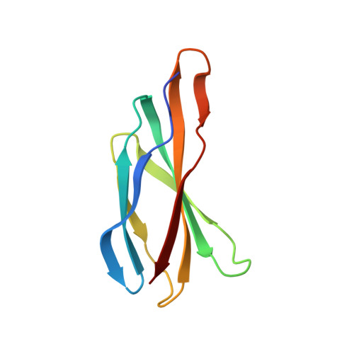
Mechanistic signs of double-barreled structure in a fluoride ion channel.

Last, N.B.Kolmakova-Partensky, L.Shane, T.Miller, C.

(2016) Elife 5

  • DOI: https://doi.org/10.7554/eLife.18767
  • Primary Citation of Related Structures:  
    5KBN, 5KOM

  • PubMed Abstract: 

    The Fluc family of F(-) ion channels protects prokaryotes and lower eukaryotes from the toxicity of environmental F(-). In bacteria, these channels are built as dual-topology dimers whereby the two subunits assemble in antiparallel transmembrane orientation. Recent crystal structures suggested that Fluc channels contain two separate ion-conduction pathways, each with two F(-) binding sites, but no functional correlates of this unusual architecture have been reported. Experiments here fill this gap by examining the consequences of mutating two conserved F(-)-coordinating phenylalanine residues. Substitution of each phenylalanine specifically extinguishes its associated F(-) binding site in crystal structures and concomitantly inhibits F(-) permeation. Functional analysis of concatemeric channels, which permit mutagenic manipulation of individual pores, show that each pore can be separately inactivated without blocking F(-) conduction through its symmetry-related twin. The results strongly support dual-pathway architecture of Fluc channels.


  • Organizational Affiliation

    Department of Biochemistry, Howard Hughes Medical Institute, Brandeis University, Waltham, United States.


Macromolecules
Find similar proteins by:  (by identity cutoff)  |  3D Structure
Entity ID: 1
MoleculeChains Sequence LengthOrganismDetailsImage
Putative fluoride ion transporter CrcB
A, B
147Escherichia coliMutation(s): 2 
Gene Names: 
Membrane Entity: Yes 
UniProt
Find proteins for Q6J5N4 (Escherichia coli)
Explore Q6J5N4 
Go to UniProtKB:  Q6J5N4
Entity Groups  
Sequence Clusters30% Identity50% Identity70% Identity90% Identity95% Identity100% Identity
UniProt GroupQ6J5N4
Sequence Annotations
Expand
  • Reference Sequence
Find similar proteins by:  (by identity cutoff)  |  3D Structure
Entity ID: 2
MoleculeChains Sequence LengthOrganismDetailsImage
monobody
C, D
97Homo sapiensMutation(s): 0 
Entity Groups  
Sequence Clusters30% Identity50% Identity70% Identity90% Identity95% Identity100% Identity
Sequence Annotations
Expand
  • Reference Sequence
Experimental Data & Validation

Experimental Data

  • Method: X-RAY DIFFRACTION
  • Resolution: 2.69 Å
  • R-Value Free: 0.230 
  • R-Value Work: 0.217 
  • R-Value Observed: 0.218 
  • Space Group: P 41
Unit Cell:
Length ( Å )Angle ( ˚ )
a = 87.5α = 90
b = 87.5β = 90
c = 144.54γ = 90
Software Package:
Software NamePurpose
MOSFLMdata reduction
Aimlessdata scaling
PHASERphasing
REFMACrefinement
PDB_EXTRACTdata extraction

Structure Validation

View Full Validation Report


Ligand Structure Quality Assessment 


Entry History & Funding Information

Deposition Data


Funding OrganizationLocationGrant Number
National Institutes of Health/National Institute of General Medical Sciences (NIH/NIGMS)United StatesRO1-GM107023

Revision History  (Full details and data files)

  • Version 1.0: 2016-08-03
    Type: Initial release
  • Version 1.1: 2016-08-10
    Changes: Database references
  • Version 1.2: 2017-09-27
    Changes: Author supporting evidence, Derived calculations
  • Version 1.3: 2019-12-25
    Changes: Author supporting evidence
  • Version 1.4: 2023-10-04
    Changes: Data collection, Database references, Derived calculations, Refinement description