5K8Y

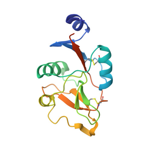
Structure of the Mus musclus Langerin carbohydrate recognition domain


Experimental Data Snapshot

  • Method: X-RAY DIFFRACTION
  • Resolution: 2.40 Å
  • R-Value Free: 0.234 
  • R-Value Work: 0.196 
  • R-Value Observed: 0.198 

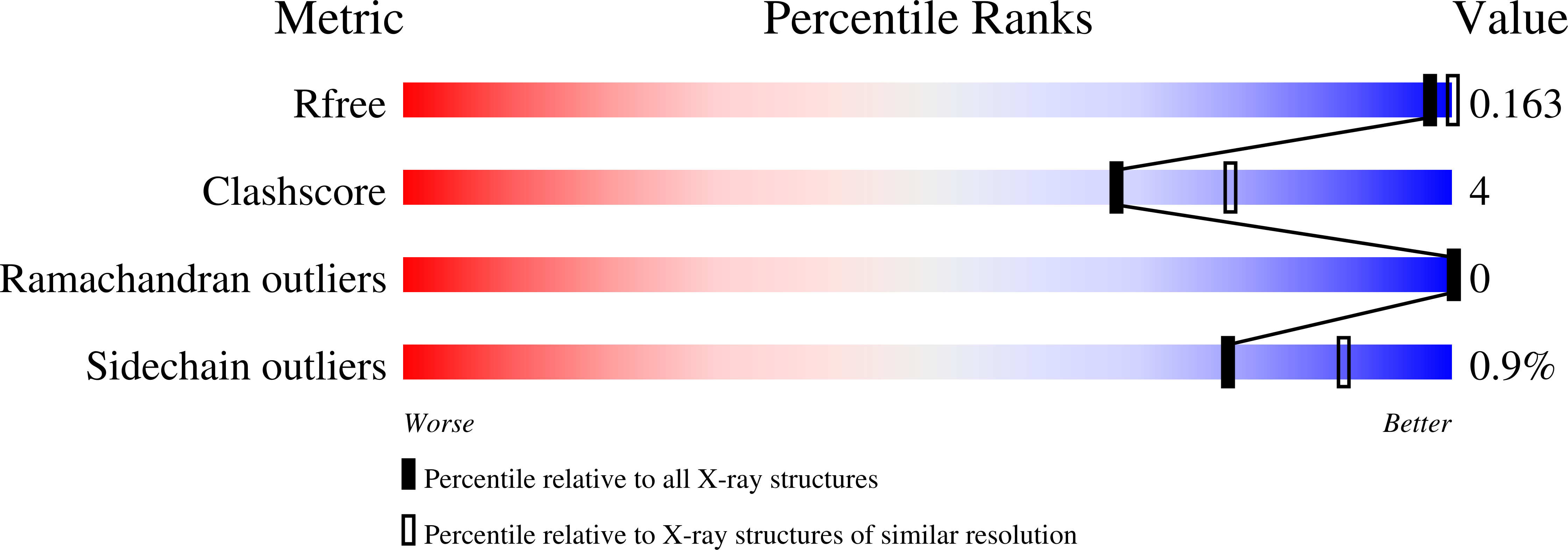
wwPDB Validation   3D Report Full Report


This is version 1.3 of the entry. See complete history


Literature

Bacterial Polysaccharide Specificity of the Pattern Recognition Receptor Langerin Is Highly Species-dependent.

Hanske, J.Schulze, J.Aretz, J.McBride, R.Loll, B.Schmidt, H.Knirel, Y.Rabsch, W.Wahl, M.C.Paulson, J.C.Rademacher, C.

(2017) J Biol Chem 292: 862-871

  • DOI: https://doi.org/10.1074/jbc.M116.751750
  • Primary Citation of Related Structures:  
    5K8Y, 5M62

  • PubMed Abstract: 

    The recognition of pathogen surface polysaccharides by glycan-binding proteins is a cornerstone of innate host defense. Many members of the C-type lectin receptor family serve as pattern recognition receptors facilitating pathogen uptake, antigen processing, and immunomodulation. Despite the high evolutionary pressure in host-pathogen interactions, it is still widely assumed that genetic homology conveys similar specificities. Here, we investigate the ligand specificities of the human and murine forms of the myeloid C-type lectin receptor langerin for simple and complex ligands augmented by structural insight into murine langerin. Although the two homologs share the same three-dimensional structure and recognize simple ligands identically, a screening of more than 300 bacterial polysaccharides revealed highly diverging avidity and selectivity for larger and more complex glycans. Structural and evolutionary conservation analysis identified a highly variable surface adjacent to the canonic binding site, potentially forming a secondary site of interaction for large glycans.


  • Organizational Affiliation

    From the Department of Biomolecular Systems, Max Planck Institute of Colloids and Interfaces, Potsdam 14424, Germany.


Macromolecules
Find similar proteins by:  (by identity cutoff)  |  3D Structure
Entity ID: 1
MoleculeChains Sequence LengthOrganismDetailsImage
C-type lectin domain family 4 member K
A, B
151Mus musculusMutation(s): 0 
Gene Names: Cd207Clec4k
UniProt
Find proteins for Q8VBX4 (Mus musculus)
Explore Q8VBX4 
Go to UniProtKB:  Q8VBX4
Entity Groups  
Sequence Clusters30% Identity50% Identity70% Identity90% Identity95% Identity100% Identity
UniProt GroupQ8VBX4
Sequence Annotations
Expand
  • Reference Sequence
Experimental Data & Validation

Experimental Data

  • Method: X-RAY DIFFRACTION
  • Resolution: 2.40 Å
  • R-Value Free: 0.234 
  • R-Value Work: 0.196 
  • R-Value Observed: 0.198 
  • Space Group: I 2 3
Unit Cell:
Length ( Å )Angle ( ˚ )
a = 143.31α = 90
b = 143.31β = 90
c = 143.31γ = 90
Software Package:
Software NamePurpose
REFMACrefinement
XDSdata reduction
XSCALEdata scaling
PHASERphasing

Structure Validation

View Full Validation Report


Entry History 

Deposition Data

Revision History  (Full details and data files)

  • Version 1.0: 2016-12-07
    Type: Initial release
  • Version 1.1: 2016-12-14
    Changes: Database references
  • Version 1.2: 2017-02-01
    Changes: Database references
  • Version 1.3: 2024-01-10
    Changes: Data collection, Database references, Derived calculations, Refinement description