5K8H

The X-ray crystal structure of a parallel poly(rA) double helix generated by rA7 at acidic pH

  • Classification: RNA
  • Organism(s): synthetic construct
  • Mutation(s): No 

  • Deposited: 2016-05-30 Released: 2016-07-06 
  • Deposition Author(s): Gleghorn, M.L., Maquat, L.E.
  • Funding Organization(s): National Institutes of Health/National Institute of General Medical Sciences (NIH/NIGMS), National Institutes of Health/National Cancer Institute (NIH/NCI)

Experimental Data Snapshot

  • Method: X-RAY DIFFRACTION
  • Resolution: 1.07 Å
  • R-Value Free: 0.108 
  • R-Value Work: 0.106 
  • R-Value Observed: 0.106 

wwPDB Validation   3D Report Full Report


This is version 1.7 of the entry. See complete history


Literature

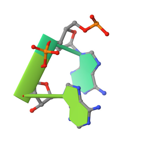
Crystal structure of a poly(rA) staggered zipper at acidic pH: evidence that adenine N1 protonation mediates parallel double helix formation.

Gleghorn, M.L.Zhao, J.Turner, D.H.Maquat, L.E.

(2016) Nucleic Acids Res 44: 8417-8424

  • DOI: https://doi.org/10.1093/nar/gkw526
  • Primary Citation of Related Structures:  
    5K8H

  • PubMed Abstract: 

    We have solved at 1. 07: Å resolution the X-ray crystal structure of a polyriboadenylic acid (poly(rA)) parallel and continuous double helix. Fifty-nine years ago, double helices of poly(rA) were first proposed to form at acidic pH. Here, we show that 7-mer oligo(rA), i.e. rA7, hybridizes and overlaps in all registers at pH 3.5 to form stacked double helices that span the crystal. Under these conditions, rA7 forms well-ordered crystals, whereas rA6 forms fragile crystalline-like structures, and rA5, rA8 and rA11 fail to crystallize. Our findings support studies from ∼50 years ago: one showed using spectroscopic methods that duplex formation at pH 4.5 largely starts with rA7 and begins to plateau with rA8; another proposed a so-called 'staggered zipper' model in which oligo(rA) strands overlap in multiple registers to extend the helical duplex. While never shown, protonation of adenines at position N1 has been hypothesized to be critical for helix formation. Bond angles in our structure suggest that N1 is protonated on the adenines of every other rAMP-rAMP helix base pair. Our data offer new insights into poly(rA) duplex formation that may be useful in developing a pH sensor.


  • Organizational Affiliation

    Department of Biochemistry and Biophysics, School of Medicine and Dentistry, University of Rochester, Rochester, NY 14642, USA Center for RNA Biology, University of Rochester, Rochester, NY 14642, USA.


Macromolecules

Find similar nucleic acids by:  Sequence   |   3D Structure  

Entity ID: 1
MoleculeChains LengthOrganismImage
RNA 7-mer7synthetic construct
Sequence Annotations
Expand
  • Reference Sequence
Small Molecules
Ligands 1 Unique
IDChains Name / Formula / InChI Key2D Diagram3D Interactions
NH4
Query on NH4

Download Ideal Coordinates CCD File 
B [auth A]AMMONIUM ION
H4 N
QGZKDVFQNNGYKY-UHFFFAOYSA-O
Experimental Data & Validation

Experimental Data

  • Method: X-RAY DIFFRACTION
  • Resolution: 1.07 Å
  • R-Value Free: 0.108 
  • R-Value Work: 0.106 
  • R-Value Observed: 0.106 
  • Space Group: P 42 21 2
Unit Cell:
Length ( Å )Angle ( ˚ )
a = 21.253α = 90
b = 21.253β = 90
c = 14.946γ = 90
Software Package:
Software NamePurpose
PHENIXrefinement
XDSdata reduction
Aimlessdata scaling
PHENIXphasing

Structure Validation

View Full Validation Report


Entry History & Funding Information

Deposition Data


Funding OrganizationLocationGrant Number
National Institutes of Health/National Institute of General Medical Sciences (NIH/NIGMS)United StatesR37 GM074593
National Institutes of Health/National Institute of General Medical Sciences (NIH/NIGMS)United StatesR01 GM22939
National Institutes of Health/National Cancer Institute (NIH/NCI)United StatesT32 CA09363

Revision History  (Full details and data files)

  • Version 1.0: 2016-07-06
    Type: Initial release
  • Version 1.1: 2016-07-13
    Changes: Database references
  • Version 1.2: 2016-07-20
    Changes: Data collection
  • Version 1.3: 2016-08-17
    Changes: Structure summary
  • Version 1.4: 2016-10-12
    Changes: Database references
  • Version 1.5: 2017-09-20
    Changes: Author supporting evidence, Database references, Derived calculations
  • Version 1.6: 2019-12-04
    Changes: Author supporting evidence
  • Version 1.7: 2023-09-27
    Changes: Data collection, Database references, Refinement description