5JZH

Cryo-EM structure of aerolysin prepore


Experimental Data Snapshot

  • Method: ELECTRON MICROSCOPY
  • Resolution: 3.90 Å
  • Aggregation State: PARTICLE 
  • Reconstruction Method: SINGLE PARTICLE 

wwPDB Validation   3D Report Full Report


This is version 1.3 of the entry. See complete history


Literature

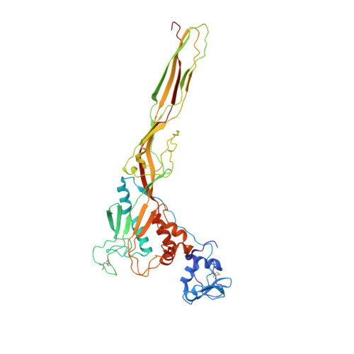
Cryo-EM structure of aerolysin variants reveals a novel protein fold and the pore-formation process.

Iacovache, I.De Carlo, S.Cirauqui, N.Dal Peraro, M.van der Goot, F.G.Zuber, B.

(2016) Nat Commun 7: 12062-12062

  • DOI: https://doi.org/10.1038/ncomms12062
  • Primary Citation of Related Structures:  
    5JZH, 5JZT, 5JZW

  • PubMed Abstract: 

    Owing to their pathogenical role and unique ability to exist both as soluble proteins and transmembrane complexes, pore-forming toxins (PFTs) have been a focus of microbiologists and structural biologists for decades. PFTs are generally secreted as water-soluble monomers and subsequently bind the membrane of target cells. Then, they assemble into circular oligomers, which undergo conformational changes that allow membrane insertion leading to pore formation and potentially cell death. Aerolysin, produced by the human pathogen Aeromonas hydrophila, is the founding member of a major PFT family found throughout all kingdoms of life. We report cryo-electron microscopy structures of three conformational intermediates and of the final aerolysin pore, jointly providing insight into the conformational changes that allow pore formation. Moreover, the structures reveal a protein fold consisting of two concentric β-barrels, tightly kept together by hydrophobic interactions. This fold suggests a basis for the prion-like ultrastability of aerolysin pore and its stoichiometry.


  • Organizational Affiliation

    Laboratory of Experimental Morphology, Institute of Anatomy, University of Bern, Baltzerstrasse 2, 3000 Bern 9, Switzerland.


Macromolecules
Find similar proteins by:  (by identity cutoff)  |  3D Structure
Entity ID: 1
MoleculeChains Sequence LengthOrganismDetailsImage
Aerolysin
A, B, C, D, E
A, B, C, D, E, F, G, H, I, J, K, L, M, N
424Aeromonas hydrophilaMutation(s): 1 
Gene Names: aerA
UniProt
Find proteins for P09167 (Aeromonas hydrophila)
Explore P09167 
Go to UniProtKB:  P09167
Entity Groups  
Sequence Clusters30% Identity50% Identity70% Identity90% Identity95% Identity100% Identity
UniProt GroupP09167
Sequence Annotations
Expand
  • Reference Sequence
Experimental Data & Validation

Experimental Data

  • Method: ELECTRON MICROSCOPY
  • Resolution: 3.90 Å
  • Aggregation State: PARTICLE 
  • Reconstruction Method: SINGLE PARTICLE 
EM Software:
TaskSoftware PackageVersion
RECONSTRUCTIONRELION1.3

Structure Validation

View Full Validation Report


Entry History & Funding Information

Deposition Data


Funding OrganizationLocationGrant Number
Swiss National Science FoundationSwitzerland--

Revision History  (Full details and data files)

  • Version 1.0: 2016-07-13
    Type: Initial release
  • Version 1.1: 2016-07-27
    Changes: Database references
  • Version 1.2: 2017-08-30
    Changes: Data collection
  • Version 1.3: 2018-10-03
    Changes: Advisory, Data collection, Derived calculations