Investigation of the mycobacterial enzyme HsaD as a potential novel target for anti-tubercular agents using a fragment-based drug design approach.
Ryan, A., Polycarpou, E., Lack, N.A., Evangelopoulos, D., Sieg, C., Halman, A., Bhakta, S., Eleftheriadou, O., McHugh, T.D., Keany, S., Lowe, E.D., Ballet, R., Abuhammad, A., Jacobs, W.R., Ciulli, A., Sim, E.(2017) Br J Pharmacol 174: 2209-2224
- PubMed: 28380256
- DOI: https://doi.org/10.1111/bph.13810
- Primary Citation of Related Structures:
5JZ9, 5JZB, 5JZS - PubMed Abstract:
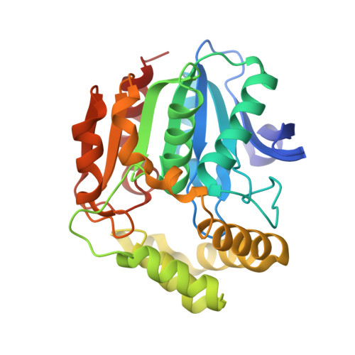
With the emergence of extensively drug-resistant tuberculosis, there is a need for new anti-tubercular drugs that work through novel mechanisms of action. The meta cleavage product hydrolase, HsaD, has been demonstrated to be critical for the survival of Mycobacterium tuberculosis in macrophages and is encoded in an operon involved in cholesterol catabolism, which is identical in M. tuberculosis and M. bovis BCG. We generated a mutant strain of M. bovis BCG with a deletion of hsaD and tested its growth on cholesterol. Using a fragment based approach, over 1000 compounds were screened by a combination of differential scanning fluorimetry, NMR spectroscopy and enzymatic assay with pure recombinant HsaD to identify potential inhibitors. We used enzymological and structural studies to investigate derivatives of the inhibitors identified and to test their effects on growth of M. bovis BCG and M. tuberculosis. The hsaD deleted strain was unable to grow on cholesterol as sole carbon source but did grow on glucose. Of seven chemically distinct 'hits' from the library, two chemical classes of fragments were found to bind in the vicinity of the active site of HsaD by X-ray crystallography. The compounds also inhibited growth of M. tuberculosis on cholesterol. The most potent inhibitor of HsaD was also found to be the best inhibitor of mycobacterial growth on cholesterol-supplemented minimal medium. We propose that HsaD is a novel therapeutic target, which should be fully exploited in order to design and discover new anti-tubercular drugs. This article is part of a themed section on Drug Metabolism and Antibiotic Resistance in Micro-organisms. To view the other articles in this section visit http://onlinelibrary.wiley.com/doi/10.1111/bph.v174.14/issuetoc.
Organizational Affiliation:
Faculty of Science, Engineering and Computing, Kingston University London, Kingston upon Thames, UK.