5JKA

Crystal structure of human JUNO (crystal form 1)


Experimental Data Snapshot

  • Method: X-RAY DIFFRACTION
  • Resolution: 2.00 Å
  • R-Value Free: 0.218 
  • R-Value Work: 0.197 
  • R-Value Observed: 0.199 

wwPDB Validation   3D Report Full Report


This is version 2.1 of the entry. See complete history


Literature

Structure of IZUMO1-JUNO reveals sperm-oocyte recognition during mammalian fertilization

Ohto, U.Ishida, H.Krayukhina, E.Uchiyama, S.Inoue, N.Shimizu, T.

(2016) Nature 534: 566-569

  • DOI: https://doi.org/10.1038/nature18596
  • Primary Citation of Related Structures:  
    5JK9, 5JKA, 5JKB, 5JKC, 5JKD, 5JKE

  • PubMed Abstract: 

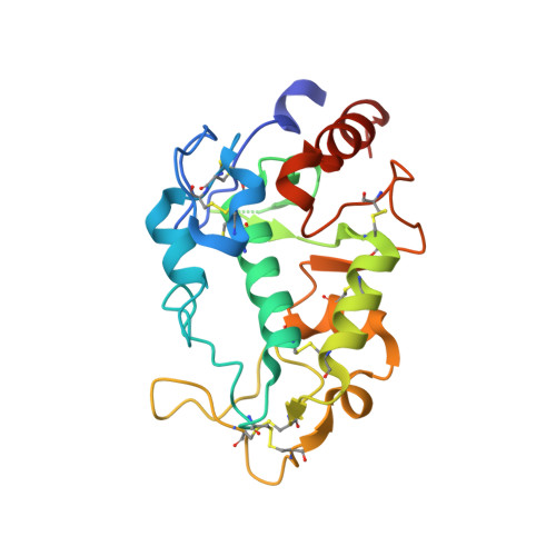
    Fertilization is a fundamental process in sexual reproduction, creating a new individual through the combination of male and female gametes. The IZUMO1 sperm membrane protein and its counterpart oocyte receptor JUNO have been identified as essential factors for sperm-oocyte interaction and fusion. However, the mechanism underlying their specific recognition remains poorly defined. Here, we show the crystal structures of human IZUMO1, JUNO and the IZUMO1-JUNO complex, establishing the structural basis for the IZUMO1-JUNO-mediated sperm-oocyte interaction. IZUMO1 exhibits an elongated rod-shaped structure comprised of a helical bundle IZUMO domain and an immunoglobulin-like domain that are each firmly anchored to an intervening β-hairpin region through conserved disulfide bonds. The central β-hairpin region of IZUMO1 provides the main platform for JUNO binding, while the surface located behind the putative JUNO ligand binding pocket is involved in IZUMO1 binding. Structure-based mutagenesis analysis confirms the biological importance of the IZUMO1-JUNO interaction. This structure provides a major step towards elucidating an essential phase of fertilization and it will contribute to the development of new therapeutic interventions for fertility, such as contraceptive agents.


  • Organizational Affiliation

    Graduate School of Pharmaceutical Sciences, The University of Tokyo, Hongo, Bunkyo-ku, Tokyo 113-0033, Japan.


Macromolecules
Find similar proteins by:  (by identity cutoff)  |  3D Structure
Entity ID: 1
MoleculeChains Sequence LengthOrganismDetailsImage
Sperm-egg fusion protein JunoA [auth B],
B [auth A]
221Homo sapiensMutation(s): 0 
Gene Names: IZUMO1RFOLR4JUNO
UniProt & NIH Common Fund Data Resources
Find proteins for A6ND01 (Homo sapiens)
Explore A6ND01 
Go to UniProtKB:  A6ND01
PHAROS:  A6ND01
GTEx:  ENSG00000183560 
Entity Groups  
Sequence Clusters30% Identity50% Identity70% Identity90% Identity95% Identity100% Identity
UniProt GroupA6ND01
Sequence Annotations
Expand
  • Reference Sequence
Oligosaccharides

Help

Entity ID: 2
MoleculeChains Length2D Diagram Glycosylation3D Interactions
alpha-D-mannopyranose-(1-3)-beta-D-mannopyranose-(1-4)-2-acetamido-2-deoxy-beta-D-glucopyranose-(1-4)-2-acetamido-2-deoxy-beta-D-glucopyranose
C
4N-Glycosylation
Glycosylation Resources
GlyTouCan:  G81315DD
GlyCosmos:  G81315DD
GlyGen:  G81315DD
Experimental Data & Validation

Experimental Data

  • Method: X-RAY DIFFRACTION
  • Resolution: 2.00 Å
  • R-Value Free: 0.218 
  • R-Value Work: 0.197 
  • R-Value Observed: 0.199 
  • Space Group: I 21 21 21
Unit Cell:
Length ( Å )Angle ( ˚ )
a = 51.92α = 90
b = 81.038β = 90
c = 235.084γ = 90
Software Package:
Software NamePurpose
REFMACrefinement
HKL-2000data reduction
HKL-2000data scaling
MOLREPphasing

Structure Validation

View Full Validation Report


Entry History 

Deposition Data

Revision History  (Full details and data files)

  • Version 1.0: 2016-06-22
    Type: Initial release
  • Version 1.1: 2016-06-29
    Changes: Database references
  • Version 1.2: 2020-02-19
    Changes: Data collection, Database references, Derived calculations
  • Version 2.0: 2020-07-29
    Type: Remediation
    Reason: Carbohydrate remediation
    Changes: Atomic model, Data collection, Derived calculations, Structure summary
  • Version 2.1: 2023-11-08
    Changes: Data collection, Database references, Derived calculations, Refinement description, Structure summary