5JGN

Spin-Labeled T4 Lysozyme Construct I9V1


Experimental Data Snapshot

  • Method: X-RAY DIFFRACTION
  • Resolution: 1.53 Å
  • R-Value Free: 0.172 
  • R-Value Work: 0.152 
  • R-Value Observed: 0.153 

wwPDB Validation   3D Report Full Report


This is version 1.2 of the entry. See complete history


Literature

Toward Precise Interpretation of DEER-Based Distance Distributions: Insights from Structural Characterization of V1 Spin-Labeled Side Chains.

Balo, A.R.Feyrer, H.Ernst, O.P.

(2016) Biochemistry 55: 5256-5263

  • DOI: https://doi.org/10.1021/acs.biochem.6b00608
  • Primary Citation of Related Structures:  
    5JGN, 5JGR, 5JGU, 5JGV, 5JGX, 5JGZ, 5KGR

  • PubMed Abstract: 

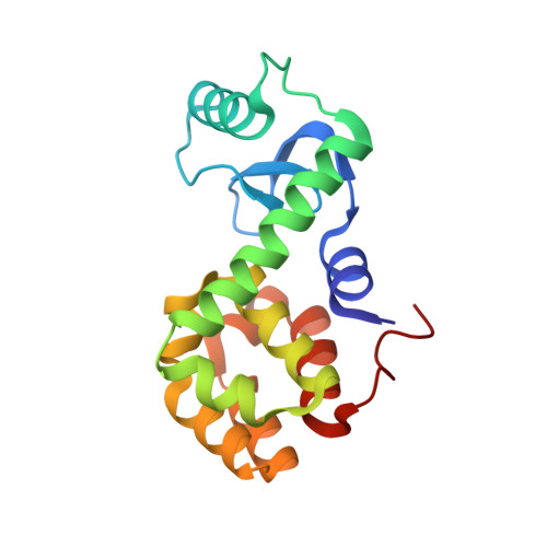
    Pulsed electron paramagnetic resonance experiments can measure individual distances between two spin-labeled side chains in proteins in the range of ∼1.5-8 nm. However, the flexibility of traditional spin-labeled side chains leads to diffuse spin density loci and thus distance distributions with relatively broad peaks, thereby complicating the interpretation of protein conformational states. Here we analyzed the spin-labeled V1 side chain, which is internally anchored and hence less flexible. Crystal structures of V1-labeled T4 lysozyme constructs carrying the V1 side chain on α-helical segments suggest that V1 side chains adopt only a few discrete rotamers. In most cases, only one rotamer is observed at a given site, explaining the frequently observed narrow distance distribution for doubly V1-labeled proteins. We used the present data to derive guidelines that may allow distance interpretation of other V1-labeled proteins for higher-precision structural modeling.


  • Organizational Affiliation

    Department of Biochemistry and ‡Department of Molecular Genetics, University of Toronto , Toronto, Ontario M5S 1A8, Canada.


Macromolecules
Find similar proteins by:  (by identity cutoff)  |  3D Structure
Entity ID: 1
MoleculeChains Sequence LengthOrganismDetailsImage
Endolysin164Escherichia phage T4Mutation(s): 3 
EC: 3.2.1.17
UniProt
Find proteins for P00720 (Enterobacteria phage T4)
Explore P00720 
Go to UniProtKB:  P00720
Entity Groups  
Sequence Clusters30% Identity50% Identity70% Identity90% Identity95% Identity100% Identity
UniProt GroupP00720
Sequence Annotations
Expand
  • Reference Sequence
Small Molecules
Ligands 4 Unique
IDChains Name / Formula / InChI Key2D Diagram3D Interactions
V1A
Query on V1A

Download Ideal Coordinates CCD File 
F [auth A]S-(1-oxyl-2,2,5,5-tetramethyl-2,5-dihydro-1H-imidazol-4-yl) methanesulfonothioate
C8 H15 N2 O3 S2
GGUBULNJTWFONN-UHFFFAOYSA-N
PO4
Query on PO4

Download Ideal Coordinates CCD File 
H [auth A]PHOSPHATE ION
O4 P
NBIIXXVUZAFLBC-UHFFFAOYSA-K
K
Query on K

Download Ideal Coordinates CCD File 
D [auth A]POTASSIUM ION
K
NPYPAHLBTDXSSS-UHFFFAOYSA-N
CL
Query on CL

Download Ideal Coordinates CCD File 
B [auth A],
C [auth A],
E [auth A],
G [auth A]
CHLORIDE ION
Cl
VEXZGXHMUGYJMC-UHFFFAOYSA-M
Experimental Data & Validation

Experimental Data

  • Method: X-RAY DIFFRACTION
  • Resolution: 1.53 Å
  • R-Value Free: 0.172 
  • R-Value Work: 0.152 
  • R-Value Observed: 0.153 
  • Space Group: P 32 2 1
Unit Cell:
Length ( Å )Angle ( ˚ )
a = 59.82α = 90
b = 59.82β = 90
c = 95.672γ = 120
Software Package:
Software NamePurpose
PHENIXrefinement
xia2data reduction
Aimlessdata scaling
PHASERphasing

Structure Validation

View Full Validation Report


Entry History & Funding Information

Deposition Data


Funding OrganizationLocationGrant Number
Canada Excellence Research ChairsCanada--

Revision History  (Full details and data files)

  • Version 1.0: 2017-02-15
    Type: Initial release
  • Version 1.1: 2017-10-25
    Changes: Database references
  • Version 1.2: 2023-09-27
    Changes: Data collection, Database references, Derived calculations, Refinement description