The Structure of LiuC, a 3-Hydroxy-3-Methylglutaconyl CoA Dehydratase Involved in Isovaleryl-CoA Biosynthesis in Myxococcus xanthus, Reveals Insights into Specificity and Catalysis.
Bock, T., Reichelt, J., Muller, R., Blankenfeldt, W.(2016) Chembiochem 17: 1658-1664
- PubMed: 27271456
- DOI: https://doi.org/10.1002/cbic.201600225
- Primary Citation of Related Structures:
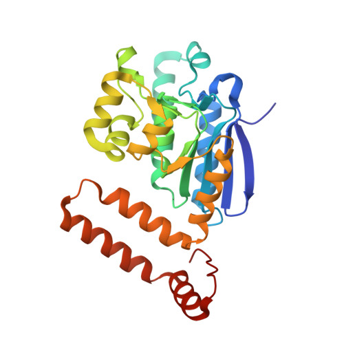
5JBW, 5JBX - PubMed Abstract:
Myxobacteria are able to produce the important metabolite isovaleryl coenzyme A by a route other than leucine degradation. The first step into this pathway is mediated by LiuC, a member of the 3-methylglutaconyl CoA hydratases (MGCH). Here we present crystal structures refined to 2.05 and 1.1 Å of LiuC in the apo form and bound to coenzyme A, respectively. By using simulated annealing we modeled the enzyme substrate complex and identified residues potentially involved in substrate binding, specificity, and catalysis. The dehydration of 3-hydroxy-3-methylglutaconyl CoA to 3-methylglutaconyl CoA catalyzed by LiuC involves Glu112 and Glu132 and likely employs the typical crotonase acid-base mechanism. In this, Tyr231 and Arg69 are key players in positioning the substrate to enable catalysis. Surprisingly, LiuC shows higher sequence and structural similarity to human MGCH than to bacterial forms, although they convert the same substrate. This study provides structural insights into the alternative isovaleryl coenzyme A biosynthesis pathway and might open a path for biofuel research, as isovaleryl-CoA is a source for isobutene, a precursor for renewable fuels and chemicals.
Organizational Affiliation:
Structure and Function of Proteins, Helmholtz Centre for Infection Research, Inhoffenstrasse 7, 38124, Braunschweig, Germany.