5J7Q

Macrophage Migration Inhibitory Factor bound to Inhibitor K664 Derivative


Experimental Data Snapshot

  • Method: X-RAY DIFFRACTION
  • Resolution: 2.05 Å
  • R-Value Free: 0.233 
  • R-Value Work: 0.195 
  • R-Value Observed: 0.199 

wwPDB Validation   3D Report Full Report


Ligand Structure Quality Assessment 


This is version 1.3 of the entry. See complete history


Literature

Irregularities in enzyme assays: The case of macrophage migration inhibitory factor.

Cisneros, J.A.Robertson, M.J.Valhondo, M.Jorgensen, W.L.

(2016) Bioorg Med Chem Lett 26: 2764-2767

  • DOI: https://doi.org/10.1016/j.bmcl.2016.04.074
  • Primary Citation of Related Structures:  
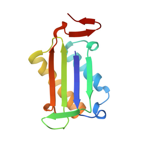
    5J7P, 5J7Q

  • PubMed Abstract: 

    Inhibitors of human macrophage migration inhibitory factor (MIF) previously reported in the literature have been reexamined by synthesis, assaying for tautomerase activity, and protein crystallography. Substantial inconsistencies between prior and current assay results are noted. They appear to arise from difficulties with the tautomerase substrates, solubility issues, and especially covalent inhibition. Incubation time variation shows that 3, 4, 6, and 9 are covalent or slow-binding inhibitors. Two protein crystal structures are provided; one confirms that the twice-discovered 3 is a covalent inhibitor.


  • Organizational Affiliation

    Department of Chemistry, Yale University, New Haven, CT 06520-8107, United States.


Macromolecules
Find similar proteins by:  (by identity cutoff)  |  3D Structure
Entity ID: 1
MoleculeChains Sequence LengthOrganismDetailsImage
Macrophage migration inhibitory factor
A, B, C
114Homo sapiensMutation(s): 0 
Gene Names: MIFGLIFMMIF
EC: 5.3.2.1 (PDB Primary Data), 5.3.3.12 (PDB Primary Data)
UniProt & NIH Common Fund Data Resources
Find proteins for P14174 (Homo sapiens)
Explore P14174 
Go to UniProtKB:  P14174
PHAROS:  P14174
GTEx:  ENSG00000240972 
Entity Groups  
Sequence Clusters30% Identity50% Identity70% Identity90% Identity95% Identity100% Identity
UniProt GroupP14174
Sequence Annotations
Expand
  • Reference Sequence
Small Molecules
Ligands 4 Unique
IDChains Name / Formula / InChI Key2D Diagram3D Interactions
6H2
Query on 6H2

Download Ideal Coordinates CCD File 
D [auth A]4-(imidazo[1,2-a]pyridin-2-yl)benzene-1,2-diol
C13 H10 N2 O2
JFYGSKNPLRBMMG-UHFFFAOYSA-N
SO4
Query on SO4

Download Ideal Coordinates CCD File 
E [auth A]
F [auth A]
G [auth B]
H [auth B]
J [auth C]
E [auth A],
F [auth A],
G [auth B],
H [auth B],
J [auth C],
K [auth C],
L [auth C]
SULFATE ION
O4 S
QAOWNCQODCNURD-UHFFFAOYSA-L
GOL
Query on GOL

Download Ideal Coordinates CCD File 
I [auth B],
N [auth C]
GLYCEROL
C3 H8 O3
PEDCQBHIVMGVHV-UHFFFAOYSA-N
IPA
Query on IPA

Download Ideal Coordinates CCD File 
M [auth C]ISOPROPYL ALCOHOL
C3 H8 O
KFZMGEQAYNKOFK-UHFFFAOYSA-N
Binding Affinity Annotations 
IDSourceBinding Affinity
6H2 Binding MOAD:  5J7Q Ki: 2.66e+4 (nM) from 1 assay(s)
BindingDB:  5J7Q Ki: 2.66e+4 (nM) from 1 assay(s)
Experimental Data & Validation

Experimental Data

  • Method: X-RAY DIFFRACTION
  • Resolution: 2.05 Å
  • R-Value Free: 0.233 
  • R-Value Work: 0.195 
  • R-Value Observed: 0.199 
  • Space Group: P 21 21 21
Unit Cell:
Length ( Å )Angle ( ˚ )
a = 67.699α = 90
b = 67.732β = 90
c = 88.152γ = 90
Software Package:
Software NamePurpose
REFMACrefinement
HKL-2000data scaling
PDB_EXTRACTdata extraction
HKL-2000data reduction
PHASERphasing

Structure Validation

View Full Validation Report


Ligand Structure Quality Assessment 


Entry History 

Deposition Data

Revision History  (Full details and data files)

  • Version 1.0: 2016-05-04
    Type: Initial release
  • Version 1.1: 2016-05-25
    Changes: Database references
  • Version 1.2: 2016-06-08
    Changes: Database references
  • Version 1.3: 2023-09-27
    Changes: Data collection, Database references, Derived calculations, Refinement description