5ILW

Crystal structure of the complex of type 1 Ribosome inactivating protein from Momordica balsamina with Uridine at 1.97 Angstrom resolution


Experimental Data Snapshot

  • Method: X-RAY DIFFRACTION
  • Resolution: 1.98 Å
  • R-Value Free: 0.214 
  • R-Value Work: 0.158 
  • R-Value Observed: 0.161 

wwPDB Validation   3D Report Full Report


Ligand Structure Quality Assessment 


This is version 1.4 of the entry. See complete history


Literature

Binding and structural studies of the complexes of type 1 ribosome inactivating protein from Momordica balsamina with uracil and uridine.

Pandey, S.N.Iqbal, N.Singh, P.K.Rastogi, N.Kaur, P.Sharma, S.Singh, T.P.

(2019) Proteins 87: 99-109

  • DOI: https://doi.org/10.1002/prot.25584
  • Primary Citation of Related Structures:  
    5ILW, 5Y48

  • PubMed Abstract: 

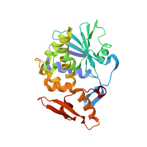
    Ribosome inactivating protein (RIP) catalyzes the cleavage of glycosidic bond formed between adenine and ribose sugar of ribosomal RNA to inactivate ribosomes. Previous structural studies have shown that RNA bases, adenine, guanine, and cytosine tend to bind to RIP in the substrate binding site. However, the mode of binding of uracil with RIP was not yet known. Here, we report crystal structures of two complexes of type 1 RIP from Momordica balsamina (MbRIP1) with base, uracil and nucleoside, uridine. The binding studies of MbRIP1 with uracil and uridine as estimated using fluorescence spectroscopy showed that the equilibrium dissociation constants (K D ) were 1.2 × 10 -6 M and 1.4 × 10 -7 M respectively. The corresponding values obtained using surface plasmon resonance (SPR) were found to be 1.4 × 10 -6 M and 1.1 × 10 -7 M, respectively. Structures of the complexes of MbRIP1 with uracil (Structure-1) and uridine (Structure-2) were determined at 1.70 and 1.98 Å resolutions respectively. Structure-1 showed that uracil bound to MbRIP1 at the substrate binding site but its mode of binding was significantly different from those of adenine, guanine and cytosine. However, the mode of binding of uridine was found to be similar to those of cytidine. As a result of binding of uracil to MbRIP1 at the substrate binding site, three water molecules were expelled while eight water molecules were expelled when uridine bound to MbRIP1.


  • Organizational Affiliation

    Department of Biophysics, All India Institute of Medical Sciences, New Delhi, India.


Macromolecules
Find similar proteins by:  (by identity cutoff)  |  3D Structure
Entity ID: 1
MoleculeChains Sequence LengthOrganismDetailsImage
Ribosome inactivating protein246Momordica balsaminaMutation(s): 0 
EC: 3.2.2.22
UniProt
Find proteins for D9J2T9 (Momordica balsamina)
Explore D9J2T9 
Go to UniProtKB:  D9J2T9
Entity Groups  
Sequence Clusters30% Identity50% Identity70% Identity90% Identity95% Identity100% Identity
UniProt GroupD9J2T9
Sequence Annotations
Expand
  • Reference Sequence
Experimental Data & Validation

Experimental Data

  • Method: X-RAY DIFFRACTION
  • Resolution: 1.98 Å
  • R-Value Free: 0.214 
  • R-Value Work: 0.158 
  • R-Value Observed: 0.161 
  • Space Group: H 3
Unit Cell:
Length ( Å )Angle ( ˚ )
a = 130.115α = 90
b = 130.115β = 90
c = 40.632γ = 120
Software Package:
Software NamePurpose
REFMACrefinement
HKL-2000data reduction
SCALEPACKdata scaling
MOLREPphasing

Structure Validation

View Full Validation Report


Ligand Structure Quality Assessment 


Entry History 

Deposition Data

Revision History  (Full details and data files)

  • Version 1.0: 2016-03-23
    Type: Initial release
  • Version 1.1: 2017-08-16
    Changes: Database references, Derived calculations
  • Version 1.2: 2019-04-03
    Changes: Data collection, Database references
  • Version 1.3: 2020-07-29
    Type: Remediation
    Reason: Carbohydrate remediation
    Changes: Data collection, Derived calculations, Structure summary
  • Version 1.4: 2023-11-08
    Changes: Data collection, Database references, Refinement description, Structure summary