5IHJ

Fusion of Maltose-binding Protein and PilA from Acinetobacter baumannii BIDMC57


Experimental Data Snapshot

  • Method: X-RAY DIFFRACTION
  • Resolution: 2.20 Å
  • R-Value Free: 0.244 
  • R-Value Work: 0.211 

wwPDB Validation   3D Report Full Report


This is version 2.0 of the entry. See complete history


Literature

Structural Diversity in the Type IV Pili of Multidrug-resistant Acinetobacter.

Piepenbrink, K.H.Lillehoj, E.Harding, C.M.Labonte, J.W.Zuo, X.Rapp, C.A.Munson, R.S.Goldblum, S.E.Feldman, M.F.Gray, J.J.Sundberg, E.J.

(2016) J Biol Chem 291: 22924-22935

  • DOI: https://doi.org/10.1074/jbc.M116.751099
  • Primary Citation of Related Structures:  
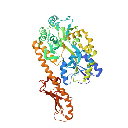
    5CFV, 5IHJ

  • PubMed Abstract: 

    Acinetobacter baumannii is a Gram-negative coccobacillus found primarily in hospital settings that has recently emerged as a source of hospital-acquired infections. A. baumannii expresses a variety of virulence factors, including type IV pili, bacterial extracellular appendages often essential for attachment to host cells. Here, we report the high resolution structures of the major pilin subunit, PilA, from three Acinetobacter strains, demonstrating that A. baumannii subsets produce morphologically distinct type IV pilin glycoproteins. We examine the consequences of this heterogeneity for protein folding and assembly as well as host-cell adhesion by Acinetobacter Comparisons of genomic and structural data with pilin proteins from other species of soil gammaproteobacteria suggest that these structural differences stem from evolutionary pressure that has resulted in three distinct classes of type IVa pilins, each found in multiple species.


  • Organizational Affiliation

    From the Institute of Human Virology and.


Macromolecules
Find similar proteins by:  (by identity cutoff)  |  3D Structure
Entity ID: 1
MoleculeChains Sequence LengthOrganismDetailsImage
Maltose-binding periplasmic protein,Fimbrial protein495Escherichia coli K-12Acinetobacter sp. 216872
This entity is chimeric
Mutation(s): 10 
Gene Names: malEb4034JW3994tfpAJ633_2062
UniProt
Find proteins for P0AEX9 (Escherichia coli (strain K12))
Explore P0AEX9 
Go to UniProtKB:  P0AEX9
Entity Groups  
Sequence Clusters30% Identity50% Identity70% Identity90% Identity95% Identity100% Identity
UniProt GroupP0AEX9
Sequence Annotations
Expand
  • Reference Sequence
Oligosaccharides

Help

Entity ID: 2
MoleculeChains Length2D Diagram Glycosylation3D Interactions
alpha-D-glucopyranose-(1-4)-alpha-D-glucopyranose-(1-4)-alpha-D-glucopyranose
B
3N/A
Glycosylation Resources
GlyTouCan:  G96370VA
GlyCosmos:  G96370VA
GlyGen:  G96370VA
Small Molecules
Biologically Interesting Molecules (External Reference) 1 Unique
Experimental Data & Validation

Experimental Data

  • Method: X-RAY DIFFRACTION
  • Resolution: 2.20 Å
  • R-Value Free: 0.244 
  • R-Value Work: 0.211 
  • Space Group: C 1 2 1
Unit Cell:
Length ( Å )Angle ( ˚ )
a = 175.07α = 90
b = 56.636β = 91.6
c = 49.997γ = 90
Software Package:
Software NamePurpose
HKL-2000data collection
HKL-2000data scaling
PHASERphasing
REFMACrefinement
PDB_EXTRACTdata extraction
HKL-2000data reduction

Structure Validation

View Full Validation Report


Entry History 

Deposition Data

Revision History  (Full details and data files)

  • Version 1.0: 2016-09-21
    Type: Initial release
  • Version 1.1: 2016-09-28
    Changes: Database references
  • Version 1.2: 2016-11-09
    Changes: Database references
  • Version 2.0: 2020-07-29
    Type: Remediation
    Reason: Carbohydrate remediation
    Changes: Atomic model, Data collection, Database references, Derived calculations, Non-polymer description, Structure summary