5I4E

Crystal Structure of Human Nonmuscle Myosin 2C motor domain


Experimental Data Snapshot

  • Method: X-RAY DIFFRACTION
  • Resolution: 2.25 Å
  • R-Value Free: 0.241 
  • R-Value Work: 0.220 
  • R-Value Observed: 0.221 

wwPDB Validation   3D Report Full Report


Ligand Structure Quality Assessment 


This is version 1.3 of the entry. See complete history


Literature

Mechanistic insights into the active site and allosteric communication pathways in human nonmuscle myosin-2C.

Chinthalapudi, K.Heissler, S.M.Preller, M.Sellers, J.R.Manstein, D.J.

(2017) Elife 6

  • DOI: https://doi.org/10.7554/eLife.32742
  • Primary Citation of Related Structures:  
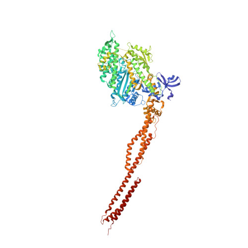
    5I4E

  • PubMed Abstract: 

    Despite a generic, highly conserved motor domain, ATP turnover kinetics and their activation by F-actin vary greatly between myosin-2 isoforms. Here, we present a 2.25 Å pre-powerstroke state (ADP⋅VO 4 ) crystal structure of the human nonmuscle myosin-2C motor domain, one of the slowest myosins characterized. In combination with integrated mutagenesis, ensemble-solution kinetics, and molecular dynamics simulation approaches, the structure reveals an allosteric communication pathway that connects the distal end of the motor domain with the active site. Disruption of this pathway by mutation of hub residue R788, which forms the center of a cluster of interactions connecting the converter, the SH1-SH2 helix, the relay helix, and the lever, abolishes nonmuscle myosin-2 specific kinetic signatures. Our results provide insights into structural changes in the myosin motor domain that are triggered upon F-actin binding and contribute critically to the mechanochemical behavior of stress fibers, actin arcs, and cortical actin-based structures.


  • Organizational Affiliation

    Institute for Biophysical Chemistry, OE4350, Hannover Medical School, Hannover, Germany.


Macromolecules
Find similar proteins by:  (by identity cutoff)  |  3D Structure
Entity ID: 1
MoleculeChains Sequence LengthOrganismDetailsImage
Myosin-14,Alpha-actinin A980Homo sapiensDictyostelium discoideum
This entity is chimeric
Mutation(s): 0 
Gene Names: MYH14KIAA2034FP17425abpAactnADDB_G0268632
UniProt & NIH Common Fund Data Resources
Find proteins for P05095 (Dictyostelium discoideum)
Explore P05095 
Go to UniProtKB:  P05095
Find proteins for Q7Z406 (Homo sapiens)
Explore Q7Z406 
Go to UniProtKB:  Q7Z406
PHAROS:  Q7Z406
GTEx:  ENSG00000105357 
Entity Groups  
Sequence Clusters30% Identity50% Identity70% Identity90% Identity95% Identity100% Identity
UniProt GroupsQ7Z406P05095
Sequence Annotations
Expand
  • Reference Sequence
Small Molecules
Ligands 2 Unique
IDChains Name / Formula / InChI Key2D Diagram3D Interactions
AOV
Query on AOV

Download Ideal Coordinates CCD File 
B [auth A]ADP ORTHOVANADATE
C10 H17 N5 O14 P2 V
SWCHWRVRYDCWAN-AZGWGOJFSA-J
MG
Query on MG

Download Ideal Coordinates CCD File 
C [auth A]MAGNESIUM ION
Mg
JLVVSXFLKOJNIY-UHFFFAOYSA-N
Experimental Data & Validation

Experimental Data

  • Method: X-RAY DIFFRACTION
  • Resolution: 2.25 Å
  • R-Value Free: 0.241 
  • R-Value Work: 0.220 
  • R-Value Observed: 0.221 
  • Space Group: P 2 21 21
Unit Cell:
Length ( Å )Angle ( ˚ )
a = 81.12α = 90
b = 125.61β = 90
c = 153.96γ = 90
Software Package:
Software NamePurpose
BUSTERrefinement
XDSdata reduction
SADABSdata scaling
PHASERphasing

Structure Validation

View Full Validation Report


Ligand Structure Quality Assessment 


Entry History & Funding Information

Deposition Data


Funding OrganizationLocationGrant Number
German Research FoundationGermanyMA 1081/21-1

Revision History  (Full details and data files)

  • Version 1.0: 2017-09-13
    Type: Initial release
  • Version 1.1: 2019-03-27
    Changes: Data collection, Database references
  • Version 1.2: 2019-04-03
    Changes: Data collection, Source and taxonomy
  • Version 1.3: 2024-01-10
    Changes: Data collection, Database references, Derived calculations, Refinement description