5I1P

Villin headpiece subdomain with a Lys30 to beta-3-homolysine substitution


Experimental Data Snapshot

  • Method: X-RAY DIFFRACTION
  • Resolution: 1.40 Å
  • R-Value Free: 0.235 
  • R-Value Work: 0.210 

wwPDB Validation   3D Report Full Report


This is version 2.0 of the entry. See complete history


Literature

Effects of Single alpha-to-beta Residue Replacements on Structure and Stability in a Small Protein: Insights from Quasiracemic Crystallization.

Kreitler, D.F.Mortenson, D.E.Forest, K.T.Gellman, S.H.

(2016) J Am Chem Soc 138: 6498-6505

  • DOI: https://doi.org/10.1021/jacs.6b01454
  • Primary Citation of Related Structures:  
    5I1N, 5I1O, 5I1P, 5I1S

  • PubMed Abstract: 

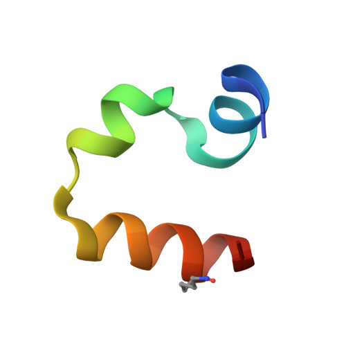
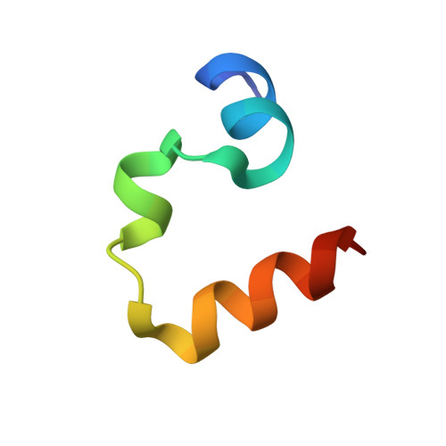
    Synthetic peptides that contain backbone modifications but nevertheless adopt folded structures similar to those of natural polypeptides are of fundamental interest and may provide a basis for biomedical applications. Such molecules can, for example, mimic the ability of natural prototypes to bind to specific target macromolecules but resist degradation by proteases. We have previously shown that oligomers containing mixtures of α- and β-amino acid residues ("α/β-peptides") can mimic the α-helix secondary structure, and that properly designed α/β-peptides can bind to proteins that evolved to bind to α-helical partners. Here we report fundamental studies that support the long-range goal of extending the α/β approach to tertiary structures. We have evaluated the impact of single α → β modifications on the structure and stability of the small and well-studied villin headpiece subdomain (VHP). The native state of this 35-residue polypeptide contains several α-helical segments packed around a small hydrophobic core. We examined α → β substitution at four solvent-exposed positions, Asn19, Trp23, Gln26 and Lys30. In each case, both the β(3) homologue of the natural α residue and a cyclic β residue were evaluated. All α → β(3) substitutions caused significant destabilization of the tertiary structure as measured by variable-temperature circular dichroism, although at some of these positions, replacing the β(3) residue with a cyclic β residue led to improved stability. Atomic-resolution structures of four VHP analogues were obtained via quasiracemic crystallization. These findings contribute to a fundamental α/β-peptide knowledge-base by confirming that β(3)-amino acid residues can serve as effective structural mimics of homologous α-amino acid residues within a natural tertiary fold, which should support rational design of functional α/β analogues of natural poly-α-peptides.


  • Organizational Affiliation

    Department of Chemistry and ‡Department of Bacteriology, University of Wisconsin-Madison , Madison, Wisconsin 53706, United States.


Macromolecules
Find similar proteins by:  (by identity cutoff)  |  3D Structure
Entity ID: 1
MoleculeChains Sequence LengthOrganismDetailsImage
Villin-1
A, B, C, D
35Gallus gallusMutation(s): 1 
UniProt
Find proteins for P02640 (Gallus gallus)
Explore P02640 
Go to UniProtKB:  P02640
Entity Groups  
Sequence Clusters30% Identity50% Identity70% Identity90% Identity95% Identity100% Identity
UniProt GroupP02640
Sequence Annotations
Expand
  • Reference Sequence
Find similar proteins by:  (by identity cutoff)  |  3D Structure
Entity ID: 2
MoleculeChains Sequence LengthOrganismDetailsImage
D-Villin headpiece subdomain
E, F, G, H
35synthetic constructMutation(s): 0 
Sequence Annotations
Expand
  • Reference Sequence
Small Molecules
Modified Residues  1 Unique
IDChains TypeFormula2D DiagramParent
B3K
Query on B3K
A, B, C, D
L-PEPTIDE LINKINGC7 H16 N2 O2LYS
Experimental Data & Validation

Experimental Data

  • Method: X-RAY DIFFRACTION
  • Resolution: 1.40 Å
  • R-Value Free: 0.235 
  • R-Value Work: 0.210 
  • Space Group: P 1
Unit Cell:
Length ( Å )Angle ( ˚ )
a = 24.57α = 96.45
b = 40.6β = 90.66
c = 59.02γ = 95.37
Software Package:
Software NamePurpose
PHENIXrefinement
XSCALEdata scaling
PHASERphasing
PDB_EXTRACTdata extraction
XDSdata reduction

Structure Validation

View Full Validation Report


Entry History & Funding Information

Deposition Data


Funding OrganizationLocationGrant Number
National Science Foundation (NSF, United States)United StatesMCB 1518160
National Institutes of Health/National Institute of General Medical Sciences (NIH/NIGMS)United StatesT32 GM00839
National Institutes of Health/National Institute of General Medical Sciences (NIH/NIGMS)United StatesR01GM061238
National Institutes of Health/National Institute of General Medical Sciences (NIH/NIGMS)United StatesT32 GM08293

Revision History  (Full details and data files)

  • Version 1.0: 2016-05-25
    Type: Initial release
  • Version 1.1: 2016-06-08
    Changes: Database references
  • Version 1.2: 2016-06-15
    Changes: Refinement description
  • Version 1.3: 2016-07-13
    Changes: Refinement description
  • Version 1.4: 2017-09-20
    Changes: Author supporting evidence, Derived calculations
  • Version 1.5: 2019-11-27
    Changes: Author supporting evidence
  • Version 1.6: 2023-09-27
    Changes: Data collection, Database references, Refinement description
  • Version 2.0: 2023-11-15
    Changes: Atomic model, Data collection