5HVQ

Alternative model of the MAGE-G1 NSE-1 complex


Experimental Data Snapshot

  • Method: X-RAY DIFFRACTION
  • Resolution: 2.92 Å
  • R-Value Free: 0.268 
  • R-Value Work: 0.237 
  • R-Value Observed: 0.238 

wwPDB Validation   3D Report Full Report


This is version 1.0 of the entry. See complete history

Re-refinement Note

This entry reflects an alternative modeling of the original data in: 3nw0


Literature

Structures of Two Melanoma-Associated Antigens Suggest Allosteric Regulation of Effector Binding.

Newman, J.A.Cooper, C.D.Roos, A.K.Aitkenhead, H.Oppermann, U.C.Cho, H.J.Osman, R.Gileadi, O.

(2016) PLoS One 11: e0148762-e0148762

  • DOI: https://doi.org/10.1371/journal.pone.0148762
  • Primary Citation of Related Structures:  
    2WA0, 4V0P, 5HVQ

  • PubMed Abstract: 

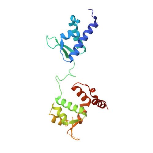
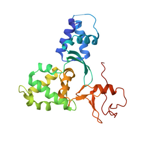
    The MAGE (melanoma associated antigen) protein family are tumour-associated proteins normally present only in reproductive tissues such as germ cells of the testis. The human genome encodes over 60 MAGE genes of which one class (containing MAGE-A3 and MAGE-A4) are exclusively expressed in tumours, making them an attractive target for the development of targeted and immunotherapeutic cancer treatments. Some MAGE proteins are thought to play an active role in driving cancer, modulating the activity of E3 ubiquitin ligases on targets related to apoptosis. Here we determined the crystal structures of MAGE-A3 and MAGE-A4. Both proteins crystallized with a terminal peptide bound in a deep cleft between two tandem-arranged winged helix domains. MAGE-A3 (but not MAGE-A4), is predominantly dimeric in solution. Comparison of MAGE-A3 and MAGE-A3 with a structure of an effector-bound MAGE-G1 suggests that a major conformational rearrangement is required for binding, and that this conformational plasticity may be targeted by allosteric binders.


  • Organizational Affiliation

    Structural Genomics Consortium, University of Oxford, ORCRB, Roosevelt Drive, Oxford, OX3 7DQ, United Kingdom.


Macromolecules
Find similar proteins by:  (by identity cutoff)  |  3D Structure
Entity ID: 1
MoleculeChains Sequence LengthOrganismDetailsImage
Melanoma-associated antigen G1A [auth D]217Homo sapiensMutation(s): 0 
Gene Names: NDNL2HCA4MAGEG1
UniProt & NIH Common Fund Data Resources
Find proteins for Q96MG7 (Homo sapiens)
Explore Q96MG7 
Go to UniProtKB:  Q96MG7
PHAROS:  Q96MG7
GTEx:  ENSG00000185115 
Entity Groups  
Sequence Clusters30% Identity50% Identity70% Identity90% Identity95% Identity100% Identity
UniProt GroupQ96MG7
Sequence Annotations
Expand
  • Reference Sequence
Find similar proteins by:  (by identity cutoff)  |  3D Structure
Entity ID: 2
MoleculeChains Sequence LengthOrganismDetailsImage
Non-structural maintenance of chromosomes element 1 homologB [auth C]238Homo sapiensMutation(s): 0 
Gene Names: NSMCE1HSPC333HSPC337
EC: 6.3.2
UniProt & NIH Common Fund Data Resources
Find proteins for Q8WV22 (Homo sapiens)
Explore Q8WV22 
Go to UniProtKB:  Q8WV22
PHAROS:  Q8WV22
GTEx:  ENSG00000169189 
Entity Groups  
Sequence Clusters30% Identity50% Identity70% Identity90% Identity95% Identity100% Identity
UniProt GroupQ8WV22
Sequence Annotations
Expand
  • Reference Sequence
Experimental Data & Validation

Experimental Data

  • Method: X-RAY DIFFRACTION
  • Resolution: 2.92 Å
  • R-Value Free: 0.268 
  • R-Value Work: 0.237 
  • R-Value Observed: 0.238 
  • Space Group: P 21 21 2
Unit Cell:
Length ( Å )Angle ( ˚ )
a = 83.257α = 90
b = 154.327β = 90
c = 53.726γ = 90
Software Package:
Software NamePurpose
PHENIXrefinement
iMOSFLMdata reduction
Aimlessdata scaling
MOLREPphasing

Structure Validation

View Full Validation Report


Entry History 

Deposition Data

Revision History  (Full details and data files)

  • Version 1.0: 2016-10-26
    Type: Initial release