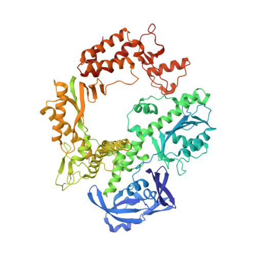
Crystal structure of Deep Vent DNA polymerase.
Hikida, Y., Kimoto, M., Hirao, I., Yokoyama, S.(2017) Biochem Biophys Res Commun 483: 52-57
- PubMed: 28063932
- DOI: https://doi.org/10.1016/j.bbrc.2017.01.007
- Primary Citation of Related Structures:
5H12 - PubMed Abstract:
DNA polymerases are useful tools in various biochemical experiments. We have focused on the DNA polymerases involved in DNA replication including the unnatural base pair between 7-(2-thienyl)imidazo[4,5-b]pyridine (Ds) and 2-nitro-4-propynylpyrrole (Px). Many reports have described the different combinations between unnatural base pairs and DNA polymerases. As an example, for the replication of the Ds-Px pair, Deep Vent DNA polymerase exhibits high efficiency and fidelity, but Taq DNA polymerase shows much lower efficiency and fidelity. In the present study, we determined the crystal structure of Deep Vent DNA polymerase in the apo form at 2.5 Å resolution. Using this structure, we constructed structural models of Deep Vent DNA polymerase complexes with DNA containing an unnatural or natural base in the replication position. The models revealed that the unnatural Ds base in the template-strand DNA clashes with the side-chain oxygen of Thr664 in Taq DNA polymerase, but not in Deep Vent DNA polymerase.
Organizational Affiliation:
RIKEN Systems and Structural Biology Center, 1-7-22 Suehiro-cho, Tsurumi, Yokohama 230-0045, Japan; Department of Biophysics and Biochemistry, Graduate School of Science, The University of Tokyo, 7-3-1 Hongo, Bunkyo-ku, Tokyo 113-0033, Japan; RIKEN Structural Biology Laboratory, 1-7-22 Suehiro-cho, Tsurumi, Yokohama 230-0045, Japan.