5GUL

Crystal structure of Tris/PPix2/Mg2+ bound form of cyclolavandulyl diphosphate synthase (CLDS) from Streptomyces sp. CL190


Experimental Data Snapshot

  • Method: X-RAY DIFFRACTION
  • Resolution: 1.73 Å
  • R-Value Free: 0.278 
  • R-Value Work: 0.216 
  • R-Value Observed: 0.219 

wwPDB Validation   3D Report Full Report


This is version 1.2 of the entry. See complete history


Literature

Structure and Mechanism of the Monoterpene Cyclolavandulyl Diphosphate Synthase that Catalyzes Consecutive Condensation and Cyclization.

Tomita, T.Kobayashi, M.Karita, Y.Yasuno, Y.Shinada, T.Nishiyama, M.Kuzuyama, T.

(2017) Angew Chem Int Ed Engl 56: 14913-14917

  • DOI: https://doi.org/10.1002/anie.201708474
  • Primary Citation of Related Structures:  
    5GUK, 5GUL

  • PubMed Abstract: 

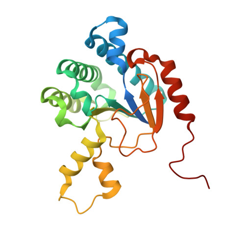
    We report the three-dimensional structure of cyclolavandulyl diphosphate (CLPP) synthase (CLDS), which consecutively catalyzes the condensation of two molecules of dimethylallyl diphosphate (DMAPP) followed by cyclization to form a cyclic monoterpene, CLPP. The structures of apo-CLDS and CLDS in complex with Tris, pyrophosphate, and Mg 2+ ion were refined at 2.00 Å resolution and 1.73 Å resolution, respectively. CLDS adopts a typical fold for cis-prenyl synthases and forms a homo-dimeric structure. An in vitro reaction using a regiospecifically 2 H-substituted DMAPP substrate revealed the intramolecular proton transfer mechanism of the CLDS reaction. The CLDS structure and structure-based mutagenesis provide mechanistic insights into this unprecedented terpene synthase. The combination of structural and mechanistic insights advances the knowledge of intricate terpene synthase-catalyzed reactions.


  • Organizational Affiliation

    Biotechnology Research Centre, The University of Tokyo, 1-1-1 Yayoi, Bunkyo-ku, Tokyo, 113-8657, Japan.


Macromolecules
Find similar proteins by:  (by identity cutoff)  |  3D Structure
Entity ID: 1
MoleculeChains Sequence LengthOrganismDetailsImage
Cyclolavandulyl diphosphate synthase
A, B, C, D
238Streptomyces sp. CL190Mutation(s): 0 
UniProt
Find proteins for X5IYJ5 (Streptomyces sp. (strain CL190))
Explore X5IYJ5 
Go to UniProtKB:  X5IYJ5
Entity Groups  
Sequence Clusters30% Identity50% Identity70% Identity90% Identity95% Identity100% Identity
UniProt GroupX5IYJ5
Sequence Annotations
Expand
  • Reference Sequence
Small Molecules
Ligands 3 Unique
IDChains Name / Formula / InChI Key2D Diagram3D Interactions
POP
Query on POP

Download Ideal Coordinates CCD File 
E [auth A]
F [auth A]
I [auth B]
J [auth B]
M [auth C]
E [auth A],
F [auth A],
I [auth B],
J [auth B],
M [auth C],
N [auth C],
Q [auth D],
R [auth D]
PYROPHOSPHATE 2-
H2 O7 P2
XPPKVPWEQAFLFU-UHFFFAOYSA-L
TRS
Query on TRS

Download Ideal Coordinates CCD File 
H [auth A],
L [auth B],
P [auth C],
T [auth D]
2-AMINO-2-HYDROXYMETHYL-PROPANE-1,3-DIOL
C4 H12 N O3
LENZDBCJOHFCAS-UHFFFAOYSA-O
MG
Query on MG

Download Ideal Coordinates CCD File 
G [auth A],
K [auth B],
O [auth C],
S [auth D]
MAGNESIUM ION
Mg
JLVVSXFLKOJNIY-UHFFFAOYSA-N
Experimental Data & Validation

Experimental Data

  • Method: X-RAY DIFFRACTION
  • Resolution: 1.73 Å
  • R-Value Free: 0.278 
  • R-Value Work: 0.216 
  • R-Value Observed: 0.219 
  • Space Group: P 1
Unit Cell:
Length ( Å )Angle ( ˚ )
a = 46.851α = 74
b = 61.661β = 84.54
c = 82.242γ = 86.04
Software Package:
Software NamePurpose
REFMACrefinement
HKL-2000data reduction
HKL-2000data scaling
PHASERphasing

Structure Validation

View Full Validation Report


Entry History & Funding Information

Deposition Data


Funding OrganizationLocationGrant Number
JSPSJapan26292058

Revision History  (Full details and data files)

  • Version 1.0: 2017-08-30
    Type: Initial release
  • Version 1.1: 2019-03-13
    Changes: Data collection, Database references
  • Version 1.2: 2023-11-08
    Changes: Data collection, Database references, Derived calculations, Refinement description