5GUJ

Crystal structure of the Bacillus subtilis DnaG RNA Polymerase Domain, natural degradation of full length DnaG


Experimental Data Snapshot

  • Method: X-RAY DIFFRACTION
  • Resolution: 2.50 Å
  • R-Value Free: 0.239 
  • R-Value Work: 0.189 
  • R-Value Observed: 0.191 

wwPDB Validation   3D Report Full Report


This is version 1.1 of the entry. See complete history


Literature

Structural Insight into the Specific DNA Template Binding to DnaG primase in Bacteria

Zhou, Y.Luo, H.Liu, Z.Yang, M.Pang, X.Sun, F.Wang, G.

(2017) Sci Rep 7: 659-659

  • DOI: https://doi.org/10.1038/s41598-017-00767-8
  • Primary Citation of Related Structures:  
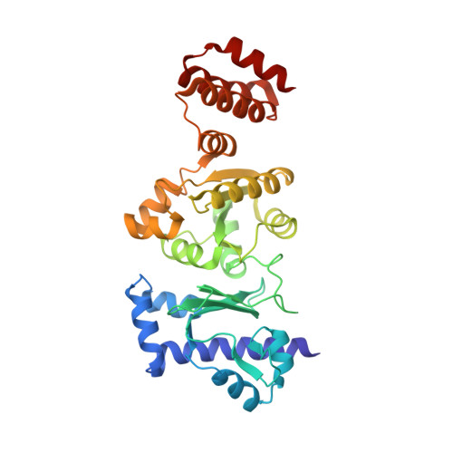
    5GUJ

  • PubMed Abstract: 

    Bacterial primase initiates the repeated synthesis of short RNA primers that are extended by DNA polymerase to synthesize Okazaki fragments on the lagging strand at replication forks. It remains unclear how the enzyme recognizes specific initiation sites. In this study, the DnaG primase from Bacillus subtilis (BsuDnaG) was characterized and the crystal structure of the RNA polymerase domain (RPD) was determined. Structural comparisons revealed that the tethered zinc binding domain plays an important role in the interactions between primase and specific template sequence. Structural and biochemical data defined the ssDNA template binding surface as an L shape, and a model for the template ssDNA binding to primase is proposed. The flexibility of the DnaG primases from B. subtilis and G. stearothermophilus were compared, and the results implied that the intrinsic flexibility of the primase may facilitate the interactions between primase and various partners in the replisome. These results shed light on the mechanism by which DnaG recognizes the specific initiation site.


  • Organizational Affiliation

    Key Laboratory of Environmental and Applied Microbiology, Chengdu Institute of Biology, Chinese Academy of Sciences, Chengdu, 610041, China.


Macromolecules
Find similar proteins by:  (by identity cutoff)  |  3D Structure
Entity ID: 1
MoleculeChains Sequence LengthOrganismDetailsImage
DNA primase324Bacillus subtilis subsp. subtilis str. 168Mutation(s): 0 
Gene Names: dnaGdnaEBSU25210
EC: 2.7.7
UniProt
Find proteins for P05096 (Bacillus subtilis (strain 168))
Explore P05096 
Go to UniProtKB:  P05096
Entity Groups  
Sequence Clusters30% Identity50% Identity70% Identity90% Identity95% Identity100% Identity
UniProt GroupP05096
Sequence Annotations
Expand
  • Reference Sequence
Experimental Data & Validation

Experimental Data

  • Method: X-RAY DIFFRACTION
  • Resolution: 2.50 Å
  • R-Value Free: 0.239 
  • R-Value Work: 0.189 
  • R-Value Observed: 0.191 
  • Space Group: P 61
Unit Cell:
Length ( Å )Angle ( ˚ )
a = 117.114α = 90
b = 117.114β = 90
c = 48.858γ = 120
Software Package:
Software NamePurpose
PHENIXrefinement
HKL-2000data reduction
HKL-2000data scaling
PHENIXphasing

Structure Validation

View Full Validation Report


Entry History 

Deposition Data

Revision History  (Full details and data files)

  • Version 1.0: 2017-04-19
    Type: Initial release
  • Version 1.1: 2023-11-08
    Changes: Data collection, Database references, Refinement description