5GU5

Crystal structure of p24gamma2 GOLD domain determined by sulfur-SAD


Experimental Data Snapshot

  • Method: X-RAY DIFFRACTION
  • Resolution: 2.80 Å
  • R-Value Free: 0.274 
  • R-Value Work: 0.224 
  • R-Value Observed: 0.226 

wwPDB Validation   3D Report Full Report


This is version 1.2 of the entry. See complete history


Literature

Crystallographic analysis of murine p24 gamma 2 Golgi dynamics domain

Nagae, M.Liebschner, D.Yamada, Y.Morita-Matsumoto, K.Matsugaki, N.Senda, T.Fujita, M.Kinoshita, T.Yamaguchi, Y.

(2017) Proteins 85: 764-770

  • DOI: https://doi.org/10.1002/prot.25242
  • Primary Citation of Related Structures:  
    5GU5

  • PubMed Abstract: 

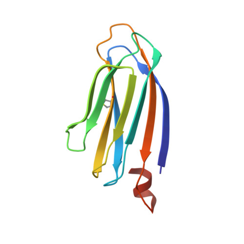
    The p24 family proteins form homo- and hetero-oligomeric complexes for efficient transport of cargo proteins from the endoplasmic reticulum to the Golgi apparatus. It consists of four subfamilies (p24α, p24β, p24γ, and p24δ). p24γ2 plays crucial roles in the selective transport of glycosylphosphatidylinositol-anchored proteins. Here, we determined the crystal structure of mouse p24γ2 Golgi dynamics (GOLD) domain at 2.8 Å resolution by the single anomalous diffraction method using intrinsic sulfur atoms. In spite of low sequence identity among p24 family proteins, p24γ2 GOLD domain assumes a β-sandwich fold, similar to that of p24β1 or p24δ1. An additional short α-helix is observed at the C-terminus of the p24γ2 GOLD domain. Intriguingly, p24γ2 GOLD domains crystallize as dimers, and dimer formation seems assisted by the short α-helix. Dimerization modes of GOLD domains are compared among p24 family proteins. Proteins 2017; 85:764-770. © 2016 Wiley Periodicals, Inc.


  • Organizational Affiliation

    Structural Glycobiology Team, Systems Glycobiology Research Group, RIKEN-Max Planck Joint Research Center, RIKEN Global Research Cluster, 2-1 Hirosawa, Wako-City, Saitama, 351-0198, Japan.


Macromolecules
Find similar proteins by:  (by identity cutoff)  |  3D Structure
Entity ID: 1
MoleculeChains Sequence LengthOrganismDetailsImage
Transmembrane emp24 domain-containing protein 5114Mus musculusMutation(s): 0 
Gene Names: Tmed5
UniProt
Find proteins for Q9CXE7 (Mus musculus)
Explore Q9CXE7 
Go to UniProtKB:  Q9CXE7
Entity Groups  
Sequence Clusters30% Identity50% Identity70% Identity90% Identity95% Identity100% Identity
UniProt GroupQ9CXE7
Sequence Annotations
Expand
  • Reference Sequence
Experimental Data & Validation

Experimental Data

  • Method: X-RAY DIFFRACTION
  • Resolution: 2.80 Å
  • R-Value Free: 0.274 
  • R-Value Work: 0.224 
  • R-Value Observed: 0.226 
  • Space Group: P 64 2 2
Unit Cell:
Length ( Å )Angle ( ˚ )
a = 79.41α = 90
b = 79.41β = 90
c = 125.948γ = 120
Software Package:
Software NamePurpose
PHENIXrefinement
HKL-2000data reduction
HKL-2000data scaling
PHENIXphasing

Structure Validation

View Full Validation Report


Entry History & Funding Information

Deposition Data


Funding OrganizationLocationGrant Number
Takeda Science FoundationJapan--
MEXTJapan25121738

Revision History  (Full details and data files)

  • Version 1.0: 2017-01-25
    Type: Initial release
  • Version 1.1: 2017-03-29
    Changes: Database references
  • Version 1.2: 2020-02-26
    Changes: Data collection